

Anastomosing hemangioma arising from the kidney: a case of slow progression in four years and review of literature
- PMID: 25973131
- PMCID: PMC4396249
Anastomosing hemangioma arising from the kidney: a case of slow progression in four years and review of literature
Abstract
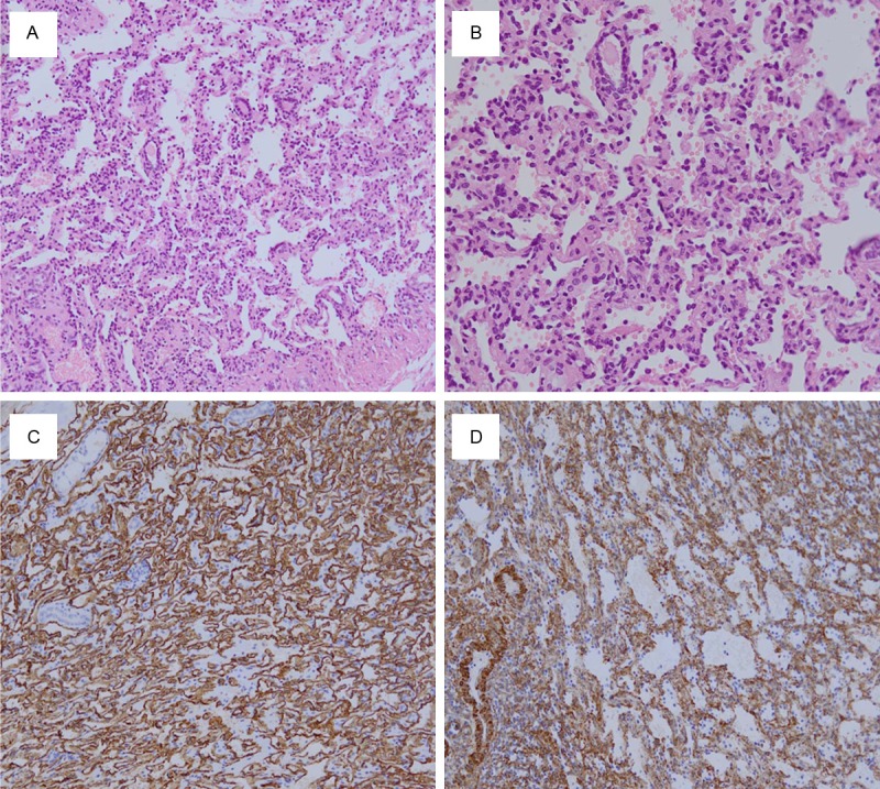
Reported herein is a renal anastomosing hemangioma which developed slowly in the past four years. A 25-year-old woman was found a mass localized in the upper portion four years ago, and only slow progression in the past four years. She underwent a laparoscopic partial nephrectomy of right kidney and diagnosed as anastomosing hemangioma. On histology the vascular components of the tumor had an anastomosing pattern without well-definite margins. Immunohistochemically, only endothelial markers (CD31, CD34) were expressed on the vascular components of tumor cells. Smooth muscle actin (SMA), cytokeratin (CK), EMA and S-100 and so on were all negative in the epithelioid tumor cells. The patient was alive at 16 months after operation, without any evidence recurrence or metastasis. Anastomosing hemangioma is an extremely rare vascular neoplasm; only 23 cases were previously described until now. Our report of anastomosing hemangioma arising from the kidney with slow progression will improve the knowledge of primary vascular tumors arising in the kidney.
Keywords: Anastomosing hemangioma; kidney; progression; rare tumors.
Figures
References
-
- Mallet R, Game X, Lefi M, Mouzin M, Malavaud B, Otal P, Joffre F, Rischmann P. [Conservative management of renal haemangioma: value of a synergistic combination of flexible ureteroscopy and CT angiography] . Prog Urol. 2007;17:108–110. - PubMed
-
- Montgomery E, Epstein JI. Anastomosing hemangioma of the genitourinary tract: a lesion mimicking angiosarcoma. Am J Surg Pathol. 2009;33:1364–1369. - PubMed
-
- Brown JG, Folpe AL, Rao P, Lazar AJ, Paner GP, Gupta R, Parakh R, Cheville JC, Amin MB. Primary vascular tumors and tumor-like lesions of the kidney: a clinicopathologic analysis of 25 cases. Am J Surg Pathol. 2010;34:942–949. - PubMed
-
- Mehta V, Ananthanarayanan V, Antic T, Krausz T, Milner J, Venkataraman G, Picken MM. Primary benign vascular tumors and tumorlike lesions of the kidney: a clinicopathologic analysis of 15 cases. Virchows Arch. 2012;461:669–676. - PubMed
-
- Kryvenko ON, Gupta NS, Meier FA, Lee MW, Epstein JI. Anastomosing hemangioma of the genitourinary system: eight cases in the kidney and ovary with immunohistochemical and ultrastructural analysis. Am J Clin Pathol. 2011;136:450–457. - PubMed
Publication types
MeSH terms
LinkOut - more resources
Full Text Sources
Medical
Research Materials
Miscellaneous