

Role of miR-100 in the radioresistance of colorectal cancer cells
- PMID: 25973296
- PMCID: PMC4396051
Role of miR-100 in the radioresistance of colorectal cancer cells
Abstract
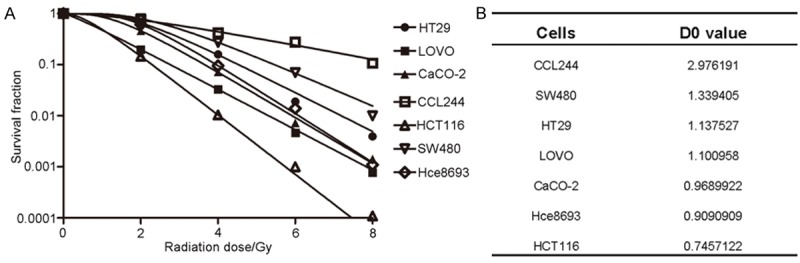
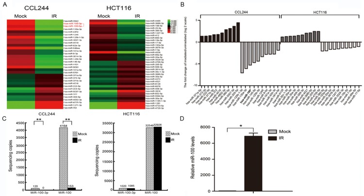
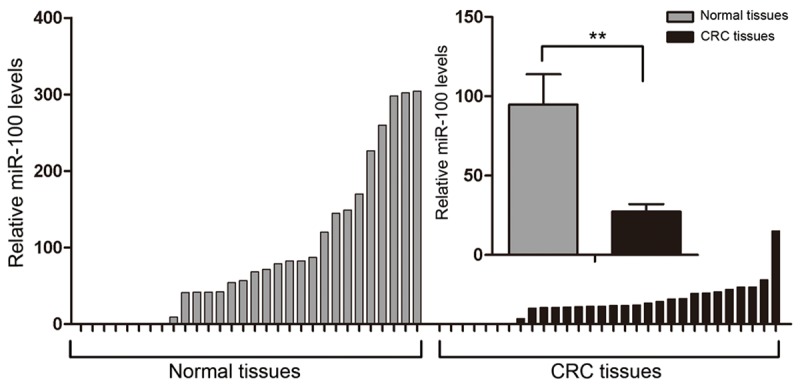
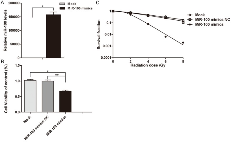
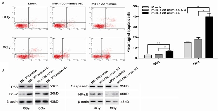
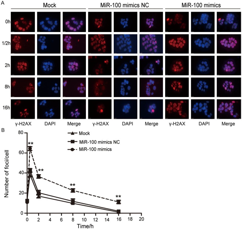
The prognosis of radioresistant colorectal cancer (CRC) is generally poor. Abnormal expression of microRNAs (miRNAs) is involved in the radiosensitivity of various tumor cells as these RNAs regulate biological signaling pathways. However, radioresistance-associated miRNAs in CRC have not yet been identified. In this study, we filtered out HCT116 and CCL-244 from seven CRC cell lines that showed the highest difference in radiosensitivity in a clonogenic assay. MiRNA sequencing identified 33 differentially expressed miRNAs (13 up-regulated and 20 down-regulated) in CCL-244 and 37 in HCT116 (20 up-regulated and 17 down-regulated) cells. MiR-100 was significantly down-regulated in CCL-244 cells after X-ray irradiation but not in HCT116 cells. Quantitative real-time PCR showed that the expression of miR-100 in CRC tissues was significantly lower than that in normal tissues. Thus, miR-100 seems to be involved in the radioresistance of CCL-244 cells. MiR-100 up-regulation sensitized CCL-244 cells to X-ray irradiation, which probably led to apoptosis and DNA double-strand breaks in these. In conclusion, to our knowledge, this is the first study to show that miR-100 may play an important role in regulating the radiosensitivity of CRC, and it may act as a new clinical target for CRC radiotherapy.
Keywords: Colorectal cancer; miR-100; miRNA profiling; radioresistance; radiosensitivity.
Figures
Similar articles
-
Insights into the radiotherapy-induced deferentially expressed RNAs in colorectal cancer management.Iran J Basic Med Sci. 2023;26(12):1380-1389. doi: 10.22038/IJBMS.2023.71259.15482. Iran J Basic Med Sci. 2023. PMID: 37970448 Free PMC article. Review.
-
MiR-7-5p/KLF4 signaling inhibits stemness and radioresistance in colorectal cancer.Cell Death Discov. 2023 Feb 2;9(1):42. doi: 10.1038/s41420-023-01339-8. Cell Death Discov. 2023. PMID: 36732504 Free PMC article.
-
LncRNA OIP5-AS1 regulates radioresistance by targeting DYRK1A through miR-369-3p in colorectal cancer cells.Eur J Cell Biol. 2018 Jun;97(5):369-378. doi: 10.1016/j.ejcb.2018.04.005. Epub 2018 Apr 14. Eur J Cell Biol. 2018. PMID: 29773344
-
Downregulation of miR-423-5p Contributes to the Radioresistance in Colorectal Cancer Cells.Front Oncol. 2021 Jan 11;10:582239. doi: 10.3389/fonc.2020.582239. eCollection 2020. Front Oncol. 2021. PMID: 33505907 Free PMC article.
-
Role of non-coding RNAs in radiosensitivity of colorectal cancer: A narrative review.Front Oncol. 2022 Jul 22;12:889658. doi: 10.3389/fonc.2022.889658. eCollection 2022. Front Oncol. 2022. PMID: 35936676 Free PMC article. Review.
Cited by
-
Downregulation of miR-1 in colorectal cancer promotes radioresistance and aggressive phenotypes.J Cancer. 2020 Jun 7;11(16):4832-4840. doi: 10.7150/jca.44753. eCollection 2020. J Cancer. 2020. PMID: 32626530 Free PMC article.
-
The Roles of Non-Coding RNAs in Radiotherapy of Gastrointestinal Carcinoma.Front Cell Dev Biol. 2022 Apr 20;10:862563. doi: 10.3389/fcell.2022.862563. eCollection 2022. Front Cell Dev Biol. 2022. PMID: 35517505 Free PMC article. Review.
-
Regulation of lnc-TLCD2-1 on Radiation Sensitivity of Colorectal Cancer and Comprehensive Analysis of Its Mechanism.Front Oncol. 2021 Jul 15;11:714159. doi: 10.3389/fonc.2021.714159. eCollection 2021. Front Oncol. 2021. PMID: 34336703 Free PMC article.
-
miR-206 enhances nasopharyngeal carcinoma radiosensitivity by targeting IGF1.Kaohsiung J Med Sci. 2017 Sep;33(9):427-432. doi: 10.1016/j.kjms.2017.05.015. Epub 2017 Jul 10. Kaohsiung J Med Sci. 2017. PMID: 28865599 Free PMC article.
-
Insights into the radiotherapy-induced deferentially expressed RNAs in colorectal cancer management.Iran J Basic Med Sci. 2023;26(12):1380-1389. doi: 10.22038/IJBMS.2023.71259.15482. Iran J Basic Med Sci. 2023. PMID: 37970448 Free PMC article. Review.
References
-
- Siegel R, Naishadham D, Jemal A. Cancer statistics, 2012. CA Cancer J Clin. 2012;62:10–29. - PubMed
-
- Engstrom PF, Arnoletti JP, Benson AB 3rd, Chen YJ, Choti MA, Cooper HS, Covey A, Dilawari RA, Early DS, Enzinger PC, Fakih MG, Fleshman J Jr, Fuchs C, Grem JL, Kiel K, Knol JA, Leong LA, Lin E, Mulcahy MF, Rao S, Ryan DP, Saltz L, Shibata D, Skibber JM, Sofocleous C, Thomas J, Venook AP, Willett C National Comprehensive Cancer Network. NCCN Clinical Practice Guidelines in Oncology: rectal cancer. J Natl Compr Canc Netw. 2009;7:838–81. - PubMed
-
- Chen G, Zhu W, Shi D, Lv L, Zhang C, Liu P, Hu W. MicroRNA-181a sensitizes human malignant glioma U87MG cells to radiation by targeting Bcl-2. Oncol Rep. 2010;23:997–1003. - PubMed
LinkOut - more resources
Full Text Sources