

Comparison of autologous (111)In-leukocytes, (18)F-FDG, (11)C-methionine, (11)C-PK11195 and (68)Ga-citrate for diagnostic nuclear imaging in a juvenile porcine haematogenous staphylococcus aureus osteomyelitis model
- PMID: 25973338
- PMCID: PMC4396013
Comparison of autologous (111)In-leukocytes, (18)F-FDG, (11)C-methionine, (11)C-PK11195 and (68)Ga-citrate for diagnostic nuclear imaging in a juvenile porcine haematogenous staphylococcus aureus osteomyelitis model
Abstract
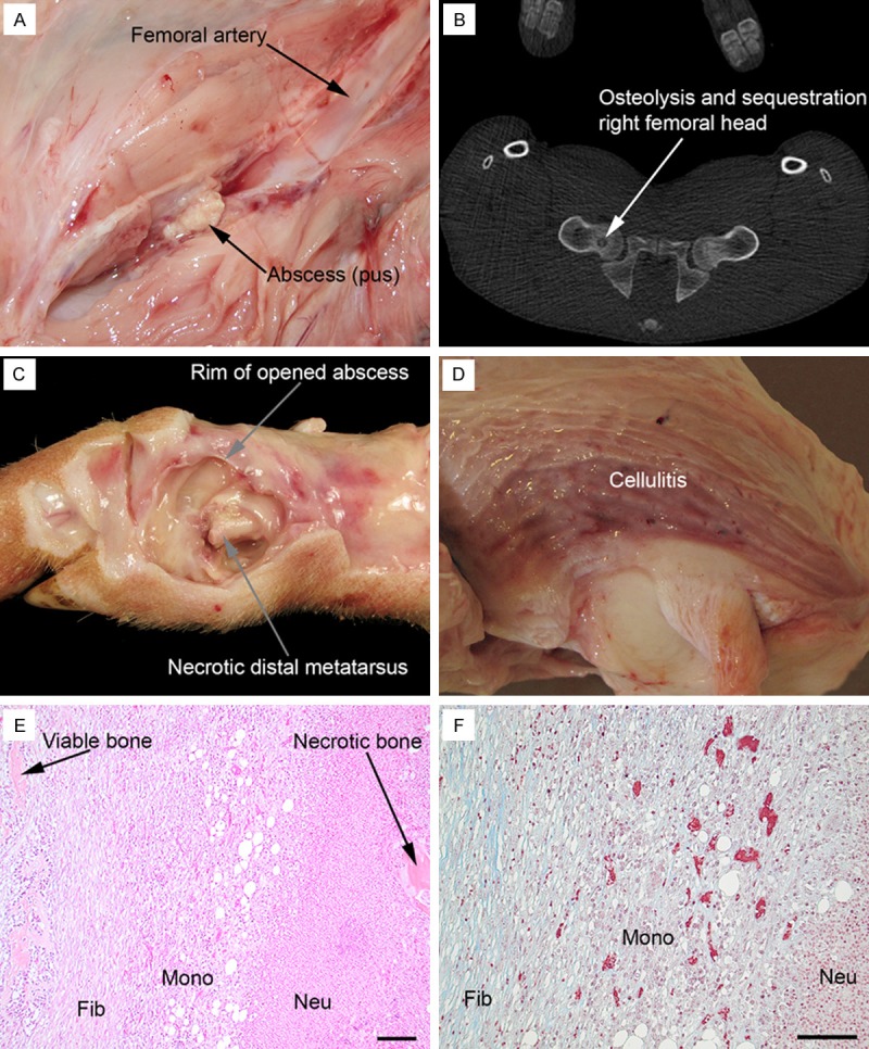
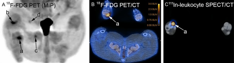
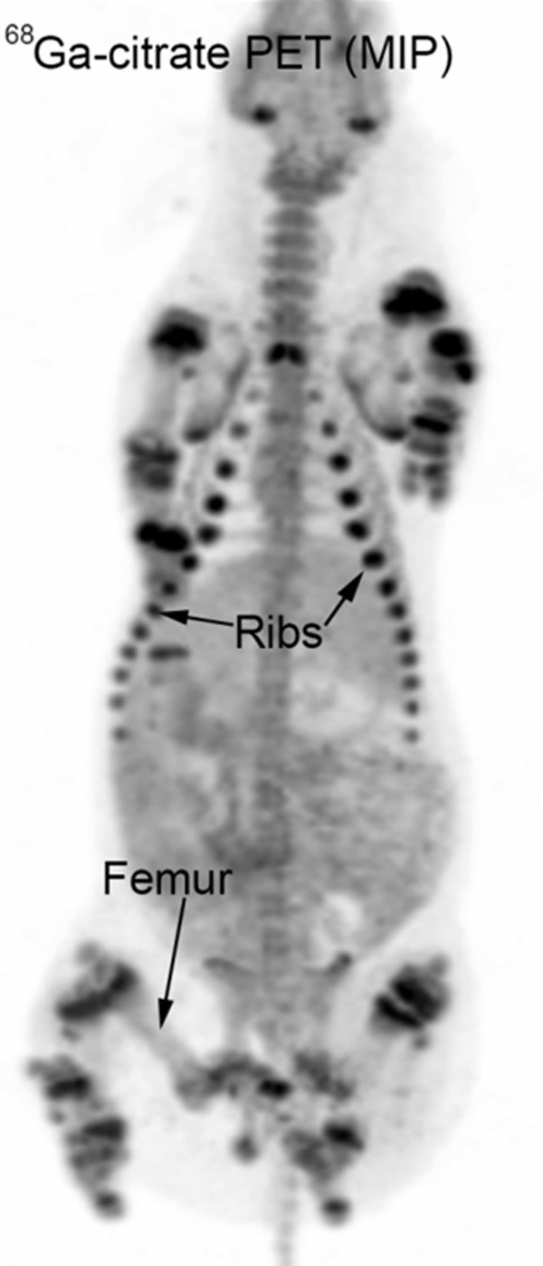
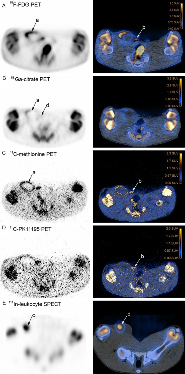
The aim of this study was to compare (111)In-labeled leukocyte single-photon emission computed tomography (SPECT) and (18)F-fluorodeoxyglucose ((18)F-FDG) positron emission tomography (PET) to PET with tracers that potentially could improve detection of osteomyelitis. We chose (11)C-methionine, (11)C-PK11195 and (68)Ga-citrate and validated their diagnostic utility in a porcine haematogenous osteomyelitis model. Four juvenile 14-15 weeks old female pigs were scanned seven days after intra-arterial inoculation in the right femoral artery with a porcine strain of Staphylococcus aureus using a sequential scan protocol with (18)F-FDG, (68)Ga-citrate, (11)C-methionine, (11)C-PK11195, (99m)Tc-Nanocoll and (111)In-labelled autologous leukocytes. This was followed by necropsy of the pigs and gross pathology, histopathology and microbial examination. The pigs developed a total of five osteomyelitis lesions, five lesions characterized as abscesses/cellulitis, arthritis in three joints and five enlarged lymph nodes. None of the tracers accumulated in joints with arthritis. By comparing the 10 infectious lesions, (18)F-FDG accumulated in nine, (111)In-leukocytes in eight, (11)C-methionine in six, (68)Ga-citrate in four and (11)C-PK11195 accumulated in only one lesion. Overall, (18)F-FDG PET was superior to (111)In-leukocyte SPECT in marking infectious and proliferative, i.e. hyperplastic, lesions. However, leukocyte SPECT was performed as early scans, approximately 6 h after injection of the leukocytes, to match the requirements of the 18 h long scan protocol. (11)C-methionine and possibly (68)Ga-citrate may be useful for diagnosis of soft issue lesions.
Keywords: Osteomyelitis; X-Ray computed; animal; domestic pigs; emission-computed; lnfection; lnflammation; models; positron-emission tomography; single-photon; staphylococcus aureus; sus scrofa; swine; tomography.
Figures
Similar articles
-
Kinetic Modelling of Infection Tracers [18F]FDG, [68Ga]Ga-Citrate, [11C]Methionine, and [11C]Donepezil in a Porcine Osteomyelitis Model.Contrast Media Mol Imaging. 2017 Oct 9;2017:9256858. doi: 10.1155/2017/9256858. eCollection 2017. Contrast Media Mol Imaging. 2017. PMID: 29114181 Free PMC article.
-
Utility of 11C-methionine and 11C-donepezil for imaging of Staphylococcus aureus induced osteomyelitis in a juvenile porcine model: comparison to autologous 111In-labelled leukocytes, 99m Tc-DPD, and 18F-FDG.Am J Nucl Med Mol Imaging. 2016 Nov 30;6(6):286-300. eCollection 2016. Am J Nucl Med Mol Imaging. 2016. PMID: 28078182 Free PMC article.
-
Biodistribution of the radionuclides (18)F-FDG, (11)C-methionine, (11)C-PK11195, and (68)Ga-citrate in domestic juvenile female pigs and morphological and molecular imaging of the tracers in hematogenously disseminated Staphylococcus aureus lesions.Am J Nucl Med Mol Imaging. 2016 Jan 28;6(1):42-58. eCollection 2016. Am J Nucl Med Mol Imaging. 2016. PMID: 27069765 Free PMC article.
-
(68)Ga-radiopharmaceuticals for PET imaging of infection and inflammation.Recent Results Cancer Res. 2013;194:189-219. doi: 10.1007/978-3-642-27994-2_11. Recent Results Cancer Res. 2013. PMID: 22918761 Review.
-
Nuclear medicine imaging of bone infections.Clin Radiol. 2016 Jul;71(7):632-46. doi: 10.1016/j.crad.2016.01.003. Epub 2016 Feb 17. Clin Radiol. 2016. PMID: 26897336 Review.
Cited by
-
Radiotracers for Bone Marrow Infection Imaging.Molecules. 2021 May 25;26(11):3159. doi: 10.3390/molecules26113159. Molecules. 2021. PMID: 34070537 Free PMC article. Review.
-
68Ga-Citrate PET of Healthy Men: Whole-Body Biodistribution Kinetics and Radiation Dose Estimates.J Nucl Med. 2022 Oct;63(10):1598-1603. doi: 10.2967/jnumed.122.263884. Epub 2022 Mar 10. J Nucl Med. 2022. PMID: 35273093 Free PMC article.
-
Choosing the right animal model for osteomyelitis research: Considerations and challenges.J Orthop Translat. 2023 Nov 29;43:47-65. doi: 10.1016/j.jot.2023.10.001. eCollection 2023 Nov. J Orthop Translat. 2023. PMID: 38094261 Free PMC article. Review.
-
Kinetic Modelling of Infection Tracers [18F]FDG, [68Ga]Ga-Citrate, [11C]Methionine, and [11C]Donepezil in a Porcine Osteomyelitis Model.Contrast Media Mol Imaging. 2017 Oct 9;2017:9256858. doi: 10.1155/2017/9256858. eCollection 2017. Contrast Media Mol Imaging. 2017. PMID: 29114181 Free PMC article.
-
Lymph Nodes Draining Infections Investigated by PET and Immunohistochemistry in a Juvenile Porcine Model.Molecules. 2022 Apr 27;27(9):2792. doi: 10.3390/molecules27092792. Molecules. 2022. PMID: 35566137 Free PMC article.
References
-
- Carek PJ, Dickerson LM, Sack JL. Diagnosis and management of osteomyelitis. Am Fam Physician. 2001;63:2413–2420. - PubMed
-
- Berbari EF, Steckelberg JM, Osmon DR. Osteomyelitis. In: Mandell GL, Bennett JE, Dolin R, editors. Mandell, Douglas, and Bennett’s Principles and Practice of Infectious Diseases. Volume 1. 6th edition. Livingstone: Elsevier Churchill; 2005. pp. 1322–1332.
-
- Lew DP, Waldvogel FA. Osteomyelitis. Lancet. 2004;364:369–379. - PubMed
-
- van der Bruggen W, Bleeker-Rovers CP, Boerman OC, Gotthardt M, Oyen WJ. PET and SPECT in osteomyelitis and prosthetic bone and joint infections: a systematic review. Semin Nucl Med. 2010;40:3–15. - PubMed
-
- Termaat MF, Raijmakers PG, Scholten HJ, Bakker FC, Patka P, Haarman HJ. The accuracy of diagnostic imaging for the assessment of chronic osteomyelitis: a systematic review and meta-analysis. J Bone Joint Surg Am. 2005;87:2464–2471. - PubMed
LinkOut - more resources
Full Text Sources