

Zebrafish Renal Pathology: Emerging Models of Acute Kidney Injury
- PMID: 25973344
- PMCID: PMC4419198
- DOI: 10.1007/s40139-015-0082-2
Zebrafish Renal Pathology: Emerging Models of Acute Kidney Injury
Abstract
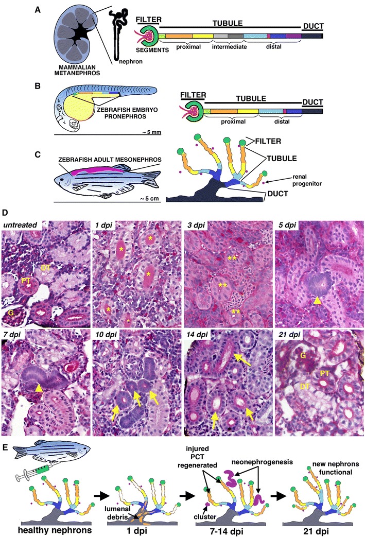
The renal system is vital to maintain homeostasis in the body, where the kidneys contain nephron functional units that remove metabolic waste from the bloodstream, regulate fluids, and balance electrolytes. Severe organ damage from toxins or ischemia that occurs abruptly can cause acute kidney injury (AKI) in which there is a rapid, life-threatening loss of these activities. Humans have a limited but poorly understood ability to regenerate damaged nephrons after AKI. However, researchers studying AKI in vertebrate animal models such as mammals, and more recently the zebrafish, have documented robust regeneration within the nephron blood filter and tubule following injury. Further, zebrafish kidneys contain progenitors that create new nephrons after AKI. Here, we review investigations in zebrafish which have established a series of exciting renal pathology paradigms that complement existing AKI models and can be implemented to discover insights into kidney regeneration and the roles of stem cells.
Keywords: Acute kidney injury; Nephron; Regeneration; Renal progenitor; Renal stem cell; Zebrafish.
Figures
Similar articles
-
Laser ablation of the zebrafish pronephros to study renal epithelial regeneration.J Vis Exp. 2011 Aug 29;(54):2845. doi: 10.3791/2845. J Vis Exp. 2011. PMID: 21897358 Free PMC article.
-
Analysis of nephron composition and function in the adult zebrafish kidney.J Vis Exp. 2014 Aug 9;(90):e51644. doi: 10.3791/51644. J Vis Exp. 2014. PMID: 25145398 Free PMC article.
-
Identification of adult nephron progenitors capable of kidney regeneration in zebrafish.Nature. 2011 Feb 3;470(7332):95-100. doi: 10.1038/nature09669. Epub 2011 Jan 26. Nature. 2011. PMID: 21270795 Free PMC article.
-
Kidney injury and regeneration in zebrafish.Semin Nephrol. 2014 Jul;34(4):437-44. doi: 10.1016/j.semnephrol.2014.06.010. Epub 2014 Jun 13. Semin Nephrol. 2014. PMID: 25217272 Review.
-
Kidney development, injury and regeneration-Zebrafish.Curr Top Dev Biol. 2025;163:307-321. doi: 10.1016/bs.ctdb.2025.01.008. Epub 2025 Feb 14. Curr Top Dev Biol. 2025. PMID: 40254347 Review.
Cited by
-
Esrrγa regulates nephron and ciliary development by controlling prostaglandin synthesis.Development. 2023 May 15;150(10):dev201411. doi: 10.1242/dev.201411. Epub 2023 May 26. Development. 2023. PMID: 37232416 Free PMC article.
-
Cannabinoid Signaling in Kidney Disease.Cells. 2023 May 18;12(10):1419. doi: 10.3390/cells12101419. Cells. 2023. PMID: 37408253 Free PMC article. Review.
-
Insights into kidney stem cell development and regeneration using zebrafish.World J Stem Cells. 2016 Feb 26;8(2):22-31. doi: 10.4252/wjsc.v8.i2.22. World J Stem Cells. 2016. PMID: 26981168 Free PMC article. Review.
-
Little fish, big catch: zebrafish as a model for kidney disease.Kidney Int. 2016 Jun;89(6):1204-10. doi: 10.1016/j.kint.2016.01.031. Epub 2016 Apr 16. Kidney Int. 2016. PMID: 27165832 Free PMC article. Review.
-
Ability of Lactobacillus brevis 47f to Alleviate the Toxic Effects of Imidacloprid Low Concentration on the Histological Parameters and Cytokine Profile of Zebrafish (Danio rerio).Int J Mol Sci. 2023 Jul 31;24(15):12290. doi: 10.3390/ijms241512290. Int J Mol Sci. 2023. PMID: 37569666 Free PMC article.
References
-
- Uchida S, Endou H. Substrate specificity to maintain cellular ATP along the mouse nephron. Am J Physiol. 1988;255(5 Pt 2):F977–F983. - PubMed
Publication types
Grants and funding
LinkOut - more resources
Full Text Sources
Other Literature Sources