. 2015 Apr 24:5:357-68.
doi: 10.1016/j.fob.2015.04.012. eCollection 2015.

Differential adaptive responses to 1- or 2-day fasting in various mouse tissues revealed by quantitative PCR analysis

Affiliations

Differential adaptive responses to 1- or 2-day fasting in various mouse tissues revealed by quantitative PCR analysis

Junya Yamamoto et al. FEBS Open Bio. .

Abstract

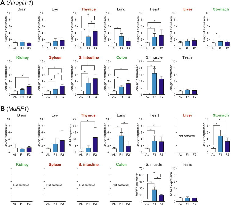
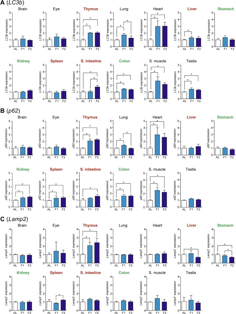
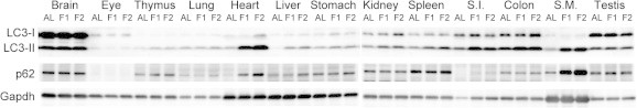
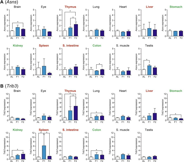
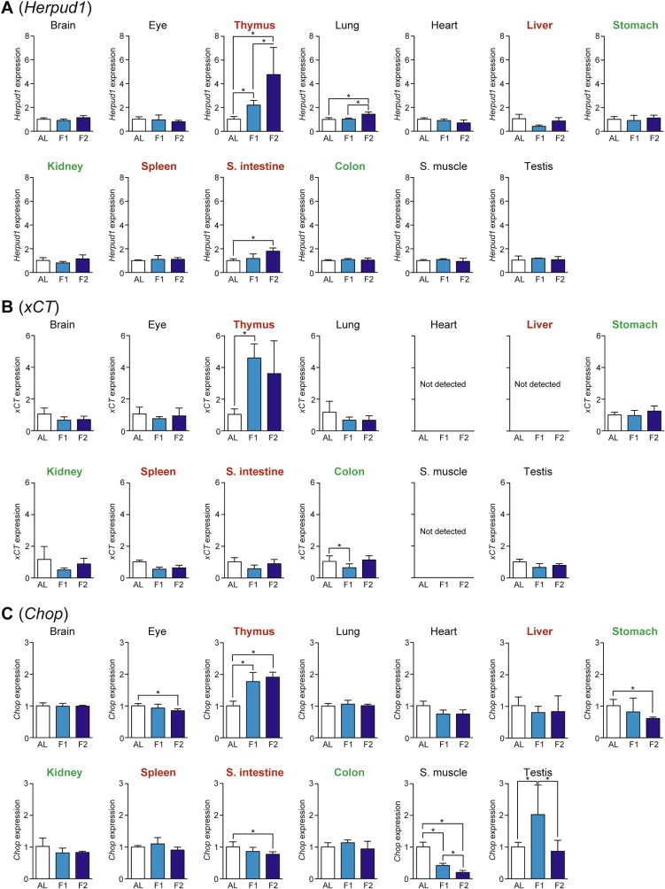
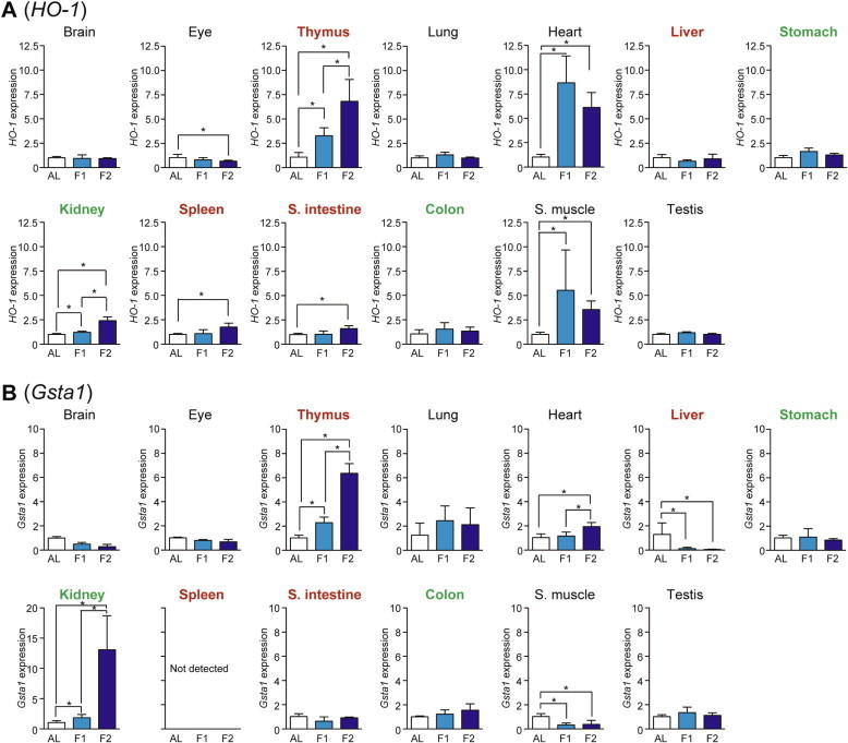
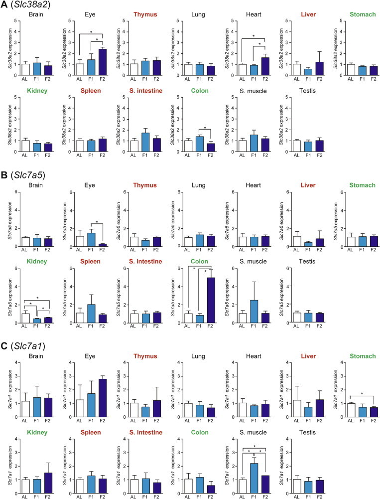
Dietary or caloric restriction confers various clinical benefits. Short-term fasting of mice is a common experimental procedure that may involve systemic metabolic remodeling, which may significantly affect experimental outputs. This study evaluated adaptive cellular responses after 1- or 2-day fasting in 13 mouse tissues by quantitative PCR using 15 marker primer sets for the activation of ubiquitin-proteasome (Atrogin-1 and MuRF1), autophagy-lysosome (LC3b, p62 and Lamp2), amino acid response (Asns, Trib3, Herpud1, xCT, and Chop), Nrf2-mediated antioxidant (HO-1 and Gsta1), and amino acid transport (Slc38a2, Slc7a5, and Slc7a1) systems. Differential activation profiles obtained in seven highly (thymus, liver, spleen, and small intestine) or mildly (stomach, kidney, and colon) atrophied tissues as well as in six non-atrophied tissues (brain, eye, lung, heart, skeletal muscle, and testis) suggested tissue-specific active metabolic remodeling.

Keywords: AL, ad libitum-fed; AST, aspartate aminotransferase; Amino acid response; Amino acid transport; Autophagy; BUN, blood urea nitrogen; CPK, creatine phosphokinase; CT, computed tomography; ER, endoplasmic reticulum; LDH, lactate dehydrogenase; Nrf2; Proteasome; TG, triglyceride; Ubiquitin; qPCR, quantitative PCR.


Figures

Fig. 1
Fig. 1
Fasting induced a rapid loss of fat but not muscle, as revealed by X-ray CT analysis. (A) Representative scanned whole body images of ad libitum-fed (AL), 1-day fasted, and 2-day fasted mice are shown. Representative lumber vertebra 1–2 (L1–L2) slice images are also shown, in which green and red regions represent fat and muscle (non-fat) depots, respectively. (B and C) Fat (B) and muscle (C) contents in the lower half of the body were estimated. Data are shown as the mean ± S.D. (n = 8), and significant changes were found in the fat tissues but not muscles; P < 0.05, ∗∗P < 0.01, ∗∗∗P < 0.001 (Mann–Whitney U-test).
Fig. 2
Fig. 2
Fasting-induced tissue-specific expression changes in genes related to the ubiquitin–proteasome system. The expression of E3 ubiquitin ligases, Atrogin-1 (A) and MuRF1 (B), in various tissues was analyzed by qPCR and normalized to Hprt expression. The expression ratios in ad libitum-fed (AL) mice were set at 1. Data are shown as the mean ± S.D. (n = 4), and the differences were significant at P < 0.05 (Mann–Whitney U-test). F1, 1-day fasting; F2, 2-day fasting; S. intestine, small intestine; S. muscle, skeletal (rectus femoris) muscle. Highly atrophied (>31% weight decrease), moderately atrophied (18–19% weight decrease), and non-atrophied tissues (no significant change) are labeled in red, green, and black, respectively.
Fig. 3
Fig. 3
Fasting-induced tissue-specific expression changes in genes related to the autophagy–lysosome system. The expression of LC3b (A), p62 (B), and Lamp2 (C) in various tissues was analyzed by qPCR and normalized to Hprt expression. The expression ratios in ad libitum-fed (AL) mice were set at 1. Data are shown as the mean ± S.D. (n = 4), and the differences were significant at P < 0.05 (Mann–Whitney U-test). F1, 1-day fasting; F2, 2-day fasting; S. intestine, small intestine; S. muscle, skeletal (rectus femoris) muscle. Highly atrophied (>31% weight decrease), moderately atrophied (18–19% weight decrease), and non-atrophied tissues (no significant change) are labeled in red, green, and black, respectively.
Fig. 4
Fig. 4
Western blot analysis of various mouse tissue homogenates for LC3b and p62 expression. Anti-LC3B antibody detected both LC3-I (∼18 kDa) and LC3-II (∼16 kDa) bands while anti-p62 and anti-GAPDH antibodies detected ∼62 kDa and ∼38 kDa bands, respectively. AL, ad libitum-fed; F1, 1-day fasting; F2, 2-day fasting; S.I., small intestine; S.M., skeletal (rectus femoris) muscle.
Fig. 5
Fig. 5
Fasting-induced tissue-specific expression changes in genes related to the amino acid response. The expression of Asns (A) and Trib3 (B) in various tissues was analyzed by qPCR and normalized to Hprt expression. The expression ratios in ad libitum-fed (AL) mice were set at 1. Data are shown as the mean ± S.D. (n = 4), and the differences were significant at P < 0.05 (Mann–Whitney U-test). F1, 1-day fasting; F2, 2-day fasting; S. intestine, small intestine; S. muscle, skeletal (rectus femoris) muscle. Highly atrophied (>31% weight decrease), moderately atrophied (18–19% weight decrease), and non-atrophied tissues (no significant change) are labeled in red, green, and black, respectively.
Fig. 6
Fig. 6
Fasting-induced tissue-specific expression changes in genes related to the amino acid response. The expression of Herpud1 (A), xCT (B), and Chop (C) in various tissues was analyzed by qPCR and normalized to Hprt expression. The expression ratios in ad libitum-fed (AL) mice were set at 1. Data are shown as the mean ± S.D. (n = 4), and the differences were significant at < 0.05 (Mann–Whitney U-test). F1, 1-day fasting; F2, 2-day fasting; S. intestine, small intestine; S. muscle, skeletal (rectus femoris) muscle. Highly atrophied (>31% weight decrease), moderately atrophied (18–19% weight decrease), and non-atrophied tissues (no significant change) are labeled in red, green, and black, respectively.
Fig. 7
Fig. 7
Fasting-induced tissue-specific expression changes in genes related to the Nrf2-mediated antioxidant system. The expression of HO-1 (A) and Gsta1 (B) in various tissues was analyzed by qPCR and normalized to Hprt expression. The expression ratios in ad libitum-fed (AL) mice were set at 1. Data are shown as the mean ± S.D. (n = 4), and the differences were significant at P < 0.05 (Mann–Whitney U-test). F1, 1-day fasting; F2, 2-day fasting; S. intestine, small intestine; S. muscle, skeletal (rectus femoris) muscle. Highly atrophied (>31% weight decrease), moderately atrophied (18–19% weight decrease), and non-atrophied tissues (no significant change) are labeled in red, green, and black, respectively.
Fig. 8
Fig. 8
Fasting-induced tissue-specific expression changes in amino acid transporter genes. The expression of Slc38a2 (A), Slc7a5 (B), and Slc7a1 (C) in various tissues was analyzed by qPCR and normalized to Hprt expression. The expression ratios in ad libitum-fed (AL) mice were set at 1. Data are shown as the mean ± S.D. (n = 4), and the differences were significant at P < 0.05 (Mann–Whitney U-test). F1, 1-day fasting; F2, 2-day fasting; S. intestine, small intestine; S. muscle, skeletal (rectus femoris) muscle. Highly atrophied (>31% weight decrease), moderately atrophied (18–19% weight decrease), and non-atrophied tissues (no significant change) are labeled in red, green, and black, respectively.

Similar articles

Cited by

References

    1. Longo V.D., Mattson M.P. Fasting: molecular mechanisms and clinical applications. Cell Metab. 2014;19:181–192. - PMC - PubMed
    1. van Praag H., Fleshner M., Schwartz M.W., Mattson M.P. Exercise, energy intake, glucose homeostasis, and the brain. J. Neurosci. 2014;34:15139–15149. - PMC - PubMed
    1. Robertson L.T., Mitchell J.R. Benefits of short-term dietary restriction in mammals. Exp. Gerontol. 2013;48:1043–1048. - PMC - PubMed
    1. Verweij M., van Ginhoven T.M., Mitchell J.R., Sluiter W., van den Engel S., Roest H.P., Torabi E., Ijzermans J.N., Hoeijmakers J.H., de Bruin R.W. Preoperative fasting protects mice against hepatic ischemia/reperfusion injury: mechanisms and effects on liver regeneration. Liver Transpl. 2011;17:695–704. - PubMed
    1. Jongbloed F., de Bruin R.W., Pennings J.L., Payan-Gomez C., van den Engel S., van Oostrom C.T., de Bruin A., Hoeijmakers J.H., van Steeg H., JN I.J., Dolle M.E. Preoperative fasting protects against renal ischemia-reperfusion injury in aged and overweight mice. PLoS One. 2014;9:e100853. - PMC - PubMed