. 2015 Feb 16;4(5):418-26.
doi: 10.1016/j.molmet.2015.02.004. eCollection 2015 May.

Munc18c mediates exocytosis of pre-docked and newcomer insulin granules underlying biphasic glucose stimulated insulin secretion in human pancreatic beta-cells

Affiliations

Munc18c mediates exocytosis of pre-docked and newcomer insulin granules underlying biphasic glucose stimulated insulin secretion in human pancreatic beta-cells

Dan Zhu et al. Mol Metab. .

Abstract

Objective: Pancreatic beta-cells express three Munc18 isoforms. Much is known about the roles of Munc18a (pre-docked secretory granules-SGs) and Munc18b (newcomer SGs and SG-SG fusion) in insulin exocytosis. Although shown to influence glucose-stimulated insulin secretion (GSIS) in rodents the precise role of Munc18c in insulin SG exocytosis has not been elucidated. We here examined the role of Munc18c in human pancreatic beta-cells.

Methods: Munc18c-shRNA/RFP lenti-virus (versus control virus) was used to knock down the expression level of Munc18c in human islets or single beta-cells. Insulin secretion and granule exocytosis were measured by performing islets perifusion, single-cell patch clamp capacitance measurements and total internal reflection fluorescence microscopy (TIRFM).

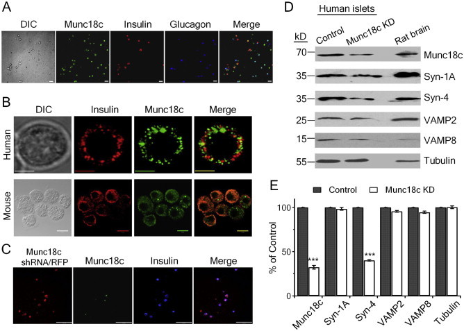
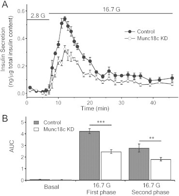
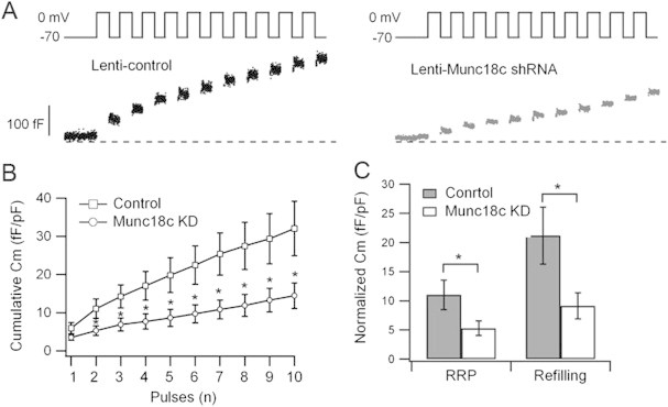
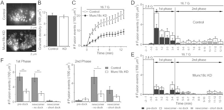
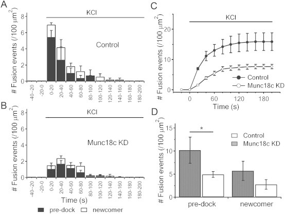
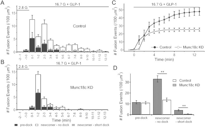
Results: Munc18c is most abundant in the cytosol of human beta-cells. Endogenous function of Munc18c was assessed by knocking down (KD) its islet expression by 70% employing lenti-shRNA virus. Munc18c-KD caused reduction in cognate syntaxin-4 islet expression but not in other exocytotic proteins, resulting in the reduction in GSIS in first- (by 42%) and second phases (by 35%). Single cell analyses of RFP-tagged Munc18c-KD beta-cells by patch clamp capacitance measurements showed inhibition in both readily-releasable pool (by 52%) and mobilization from the reserve pool (by 57%). TIRFM to assess single SG behavior showed that Munc18c-KD inhibition of first phase GSIS was attributed to reduction in exocytosis of pre-docked and newcomer SGs, and second phase inhibition attributed entirely to reduction in newcomer SG fusion (SGs that undergo minimal residence or docking time at the plasma membrane before fusion).

Conclusion: Munc18c is involved in the distinct molecular machineries that affect exocytosis of both predocked and newcomer SG pools that underlie Munc18c's role in first and second phases of GSIS, respectively.

Keywords: Ad, adenovirus; CmPatch, clamp capacitance measurements; EGFP, enhanced green fluorescent protein; Exocytosis; GLP-1, glucagon-like peptide-1; GSIS, glucose-stimulated insulin secretion; Human islets; KD, knock down; Munc18c; NPY, neuropeptide Y; Newcomer insulin granules; PM, plasma membrane; RRP, readily releasable pool; SG, secretory insulin-containing granule; SM, Sec1/Munc18-like protein; SNAP25/23, synaptosomal-associated protein of 25/23 kD; SNARE, soluble N-ethylmaleimide-sensitive factor attachment protein receptor; Syn, syntaxin; T2DM, type 2 diabetes mellitus; TIRFM, total internal reflection fluorescence microscopy; VAMPs, Vesicle Associated Membrane Proteins; t-, target-; v-, vesicle-.


Figures

Figure 1
Figure 1
Munc18c is present in human islet beta-cells and alpha-cells and its expression is depleted by lenti-shRNA. (A) Representative confocal images show Munc18c (green) is present in both insulin-containing beta-cells (red) and glucagon-containing alpha-cells (blue). Scale bars: 100 μm. (B) Representative confocal images show Munc18c is abundant in the cell cytosol of human (top images, scale bars: 5 μm) and C57BL/6 mouse (bottom images; scale bars: 10 μm) beta-cells. (C) Triple labeling of lenti-Munc18c-shRNA/RFP, Munc18c/FITC and insulin/Cy5 in dispersed human islet cells. Scale bars: 100 μm. (D–E) Western blotting analysis of lenti-Munc18c-shRNA/RFP-induced knockdown of Munc18c expression in human islets. Representative blots of three separate experiments, where means ± SEMs are shown in (E).
Figure 2
Figure 2
Depletion of Munc18c in human islets causes reduction in first- and second-phase GSIS. (A) Human islets infected with lenti-Munc18c-shRNA/RFP or lenti-/RFP (control) were subjected to islet perifusion assays. (B) Area under the curve (AUC) analysis of first-phase (8–21 min), second-phase (22–46 min) of GSIS. Basal secretion was not significantly affected. **p < 0.01; ***p < 0.001; N = 6.
Figure 3
Figure 3
Depletion of Munc18c in human beta-cells causes reduction in RRP and SG pool mobilization. Patch clamp Cm performed on single human beta-cells (RFP-positive) infected with lenti-control shRNA/RFP or lenti-Munc18c-shRNA/RFP. (A) Representative recordings of exocytosis during a train of 500 ms depolarizations from −70 to 0 mV. (B) Cumulative changes in cell capacitance normalized to basal cell membrane capacitance (fF/pF) in control (n = 11 cells) and Munc18c-KD (n = 12 cells) beta-cells. (C) Size of RRP (ΔCm1st–2nd pulse) and rate of SG mobilization (ΔCm3rd–10th pulse). Values represent means ± SEMs. *p < 0.05.
Figure 4
Figure 4
Munc18c depletion inhibits biphasic GSIS by reducing pre-docked and no-docked newcomer SGs exocytosis. (A) TIRF images of docked insulin SGs in control (top) or Munc18c-KD (bottom) human islet beta-cells. Scale bars, 2 μm. (B) Comparison of averaged SG densities before stimulation. (C) Normalized cumulative fusion events of SGs per unit area from control and Munc18c-KD beta-cells. (D,E) Histograms of fusion events in first-phase (first 4 min stimulation) and second-phase (5–13 min) showing insulin SG exocytosis dynamics caused by 16.7 mM glucose stimulation from lenti-control/RFP (D) and lenti-Munc18c-shRNA/RFP (E) -treated human beta-cells. Black, white and grey bars indicate pre-docked, no-dock and short-dock newcomer SGs, respectively; Control: 12 cells; Munc18c-KD: 11 cells, expressed as means ± SEMs. (F) Summary of the three modes of fusion events in first (left) and second phases (right), shown as means ± SEMs; *p < 0.05; **p < 0.01.
Figure 5
Figure 5
Munc18c depletion diminishes high-K+-evoked exocytosis of previously-docked insulin SGs. (A,B) Insulin SG exocytosis dynamics evoked by 50 mmol/l KCl from control (A) and Munc18c-KD (B) human beta-cells. Black and white bars indicate pre-docked and newcomer SGs, respectively. Data from 10 cells for each group, expressed as means ± SEMs. (C) Normalized cumulative fusion events of SGs per unit area from control (black circles) and Munc18c-KD (white circles) beta-cells. (D) Summary of fusion events from pre-docked and newcomer SGs in first- and second-phase after 50 mmol/l glucose stimulation. Values represent means ± SEMs. *p < 0.05.
Figure 6
Figure 6
GLP-1-potentiated GSIS rescues only pre-docked SGs exocytosis. (A,B) Insulin SG exocytosis dynamics caused by 16.7 mmol/l glucose and 10 nmol/l GLP-1 from lenti-control/RFP and lenti-Munc18c-shRNA/RFP-treated human beta-cells. Data obtained from 10 cells for each, and expressed as means ± SEMs. (C) Normalized cumulative fusion events of insulin SGs per unit area from control (black circles) and Munc18c-KD (white circles) human beta-cells. (D) Summary of fusion events from pre-docked SGs and newcomer SGs after 16.7 mmol/l glucose and 10 nmol/l GLP-1 stimulation. Values represent the means ± SEMs. **p < 0.01.

Similar articles

Cited by

References

    1. Sudhof T.C., Rothman J.E. Membrane fusion: grappling with SNARE and SM proteins. Science. 2009;323(5913):474–477. - PMC - PubMed
    1. Jahn R., Scheller R.H. SNAREs–engines for membrane fusion. Nature Reviews Molecular Cell Biology. 2006;7(9):631–643. - PubMed
    1. Garcia E.P., Gatti E., Butler M., Burton J., De Camilli P. A rat brain Sec1 homologue related to Rop and UNC18 interacts with syntaxin. Proceedings of the National Academy of Sciences of the United States of America. 1994;91(6):2003–2007. - PMC - PubMed
    1. Tamori Y., Kawanishi M., Niki T., Shinoda H., Araki S., Okazawa H. Inhibition of insulin-induced GLUT4 translocation by Munc18c through interaction with syntaxin4 in 3T3-L1 adipocytes. The Journal of Biological Chemistry. 1998;273(31):19740–19746. - PubMed
    1. Tellam J.T., McIntosh S., James D.E. Molecular identification of two novel Munc-18 isoforms expressed in non-neuronal tissues. The Journal of Biological Chemistry. 1995;270(11):5857–5863. - PubMed

LinkOut - more resources