

Identification of Sestrin3 Involved in the In vitro Resistance of Colorectal Cancer Cells to Irinotecan
- PMID: 25973791
- PMCID: PMC4431826
- DOI: 10.1371/journal.pone.0126830
Identification of Sestrin3 Involved in the In vitro Resistance of Colorectal Cancer Cells to Irinotecan
Abstract
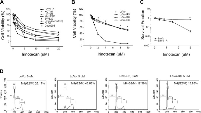
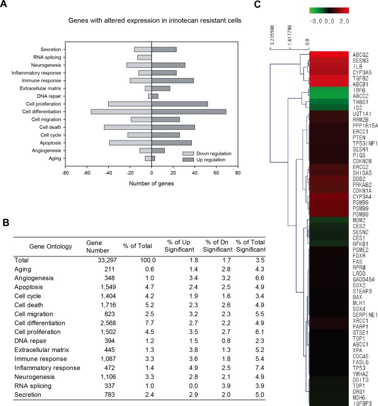
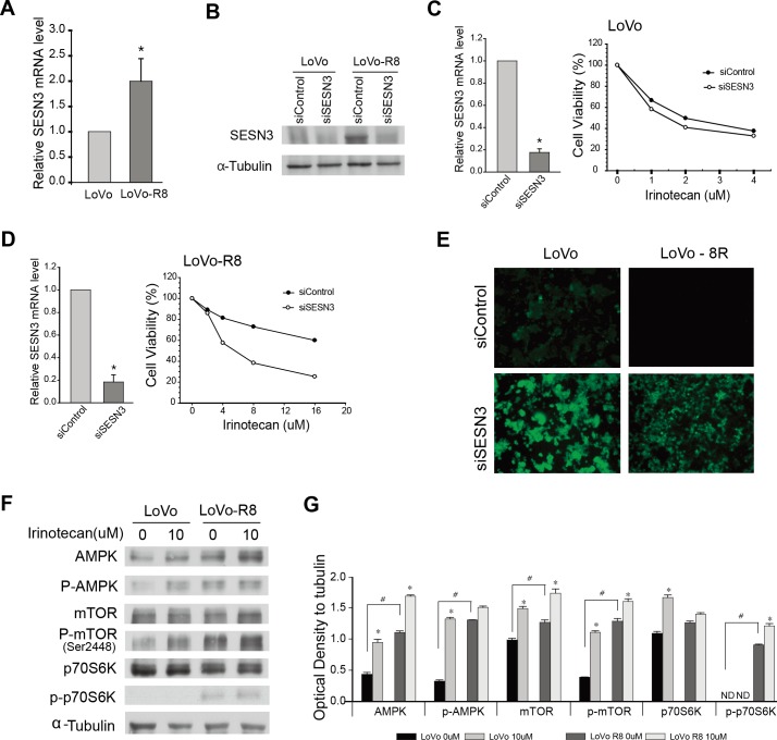
Irinotecan, an analogue of camptothecin, is frequently used as a single agent or in combination with other anticancer drugs for the treatment of colorectal cancer. However, the drug resistance of tumors is a major obstacle to successful cancer treatment. In this study, we established that cells acquire chronic resistance to irinotecan. We profiled their differential gene expression using microarray. After gene ontology (GO) and KEGG pathway analysis of the microarray data, we specifically investigated whether Sestrin3 could decrease irinotecan resistance. Our results revealed that Sestrin3 enhanced the anticancer effect of irinotecan in vitro in LoVo cells that had acquired resistance to irinotecan. Irinotecan-resistant LoVo cells showed lower reactive oxygen species (ROS) production than their irinotecan-sensitive parental cells. ROS production was increased by Sestrin3 knockdown in irinotecan-resistant LoVo cells. Our results indicate that Sestrin3 might be a good target to develop therapeutics that can help to overcome resistance to irinotecan.
Conflict of interest statement
Figures
Similar articles
-
Curcumin enhances the effects of irinotecan on colorectal cancer cells through the generation of reactive oxygen species and activation of the endoplasmic reticulum stress pathway.Oncotarget. 2017 Jun 20;8(25):40264-40275. doi: 10.18632/oncotarget.16828. Oncotarget. 2017. PMID: 28402965 Free PMC article.
-
Heat shock protein 27 is associated with irinotecan resistance in human colorectal cancer cells.FEBS Lett. 2007 Apr 17;581(8):1649-56. doi: 10.1016/j.febslet.2007.02.075. Epub 2007 Mar 22. FEBS Lett. 2007. PMID: 17395183
-
RhoA regulates resistance to irinotecan by regulating membrane transporter and apoptosis signaling in colorectal cancer.Oncotarget. 2016 Dec 27;7(52):87136-87146. doi: 10.18632/oncotarget.13548. Oncotarget. 2016. PMID: 27888624 Free PMC article.
-
Irinotecan in the first-line treatment of colorectal cancer.Oncology (Williston Park). 1998 Aug;12(8 Suppl 6):54-8. Oncology (Williston Park). 1998. PMID: 9726092 Review.
-
[Second-line irinotecan chemotherapy in the treatment of metastatic colorectal cancers: phase III trials].Bull Cancer. 1998 Dec;Spec No:38-42. Bull Cancer. 1998. PMID: 9932083 Review. French.
Cited by
-
The functions and roles of sestrins in regulating human diseases.Cell Mol Biol Lett. 2022 Jan 3;27(1):2. doi: 10.1186/s11658-021-00302-8. Cell Mol Biol Lett. 2022. PMID: 34979914 Free PMC article. Review.
-
Gene promoter and exon DNA methylation changes in colon cancer development - mRNA expression and tumor mutation alterations.BMC Cancer. 2018 Jun 27;18(1):695. doi: 10.1186/s12885-018-4609-x. BMC Cancer. 2018. PMID: 29945573 Free PMC article.
References
-
- Cunningham D, Atkin W, Lenz HJ, Lynch HT, Minsky B, Nordlinger B, et al. Colorectal cancer. Lancet. 2010;375(9719):1030–47. doi: 10.1016/S0140-6736(10)60353-4 . - DOI - PubMed
-
- Mayer RJ. Targeted therapy for advanced colorectal cancer—more is not always better. The New England journal of medicine. 2009;360(6):623–5. doi: 10.1056/NEJMe0809343 . - DOI - PubMed
-
- Wolpin BM, Mayer RJ. Systemic treatment of colorectal cancer. Gastroenterology. 2008;134(5):1296–310. doi: 10.1053/j.gastro.2008.02.098 ; PubMed Central PMCID: PMC2528832. - DOI - PMC - PubMed
-
- Xu Y, Villalona-Calero MA. Irinotecan: mechanisms of tumor resistance and novel strategies for modulating its activity. Ann Oncol. 2002;13(12):1841–51. . - PubMed
-
- Budanov AV, Sablina AA, Feinstein E, Koonin EV, Chumakov PM. Regeneration of peroxiredoxins by p53-regulated sestrins, homologs of bacterial AhpD. Science. 2004;304(5670):596–600. doi: 10.1126/science.1095569 . - DOI - PubMed
Publication types
MeSH terms
Substances
Associated data
- Actions
LinkOut - more resources
Full Text Sources
Other Literature Sources
Molecular Biology Databases
