

Integrin α4 Enhances Metastasis and May Be Associated with Poor Prognosis in MYCN-low Neuroblastoma
- PMID: 25973900
- PMCID: PMC4431816
- DOI: 10.1371/journal.pone.0120815
Integrin α4 Enhances Metastasis and May Be Associated with Poor Prognosis in MYCN-low Neuroblastoma
Abstract
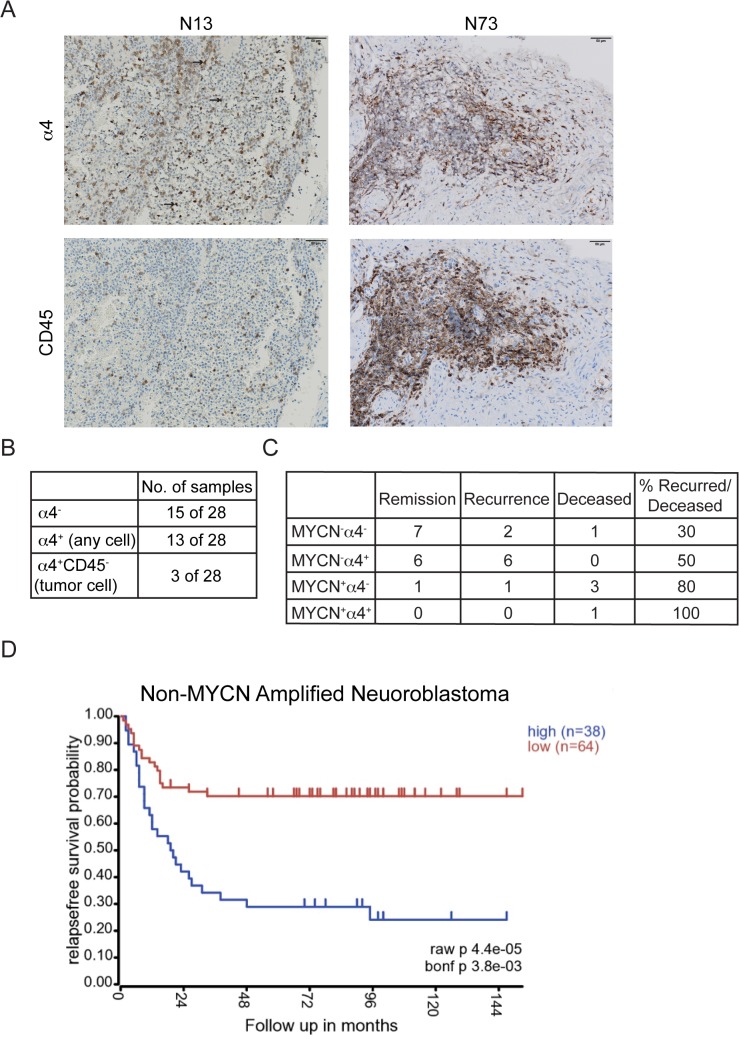
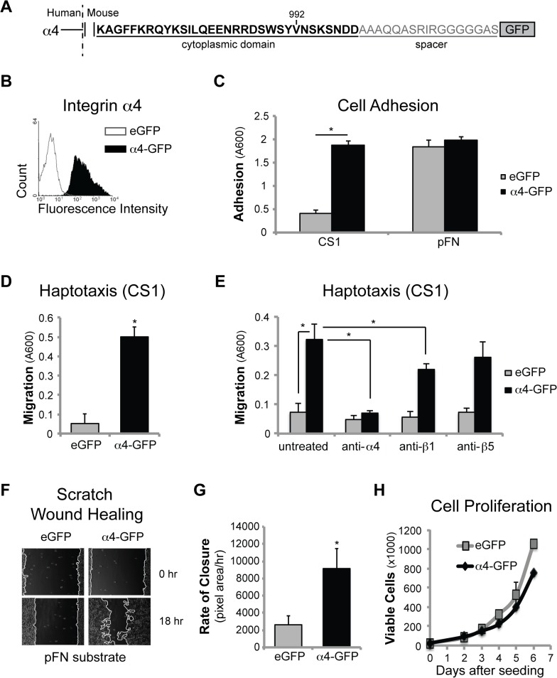
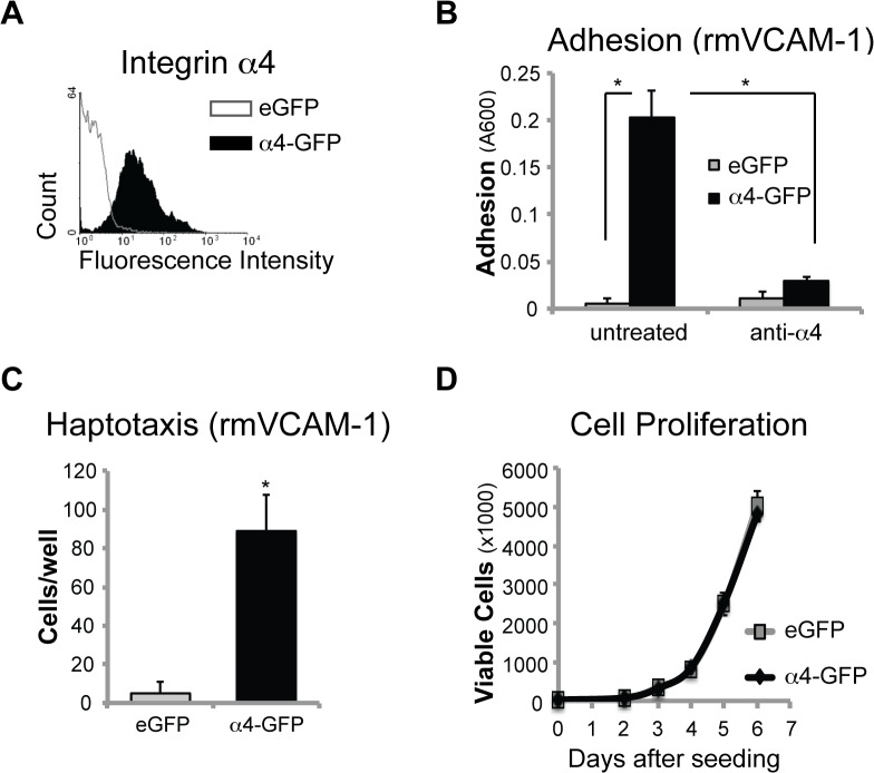
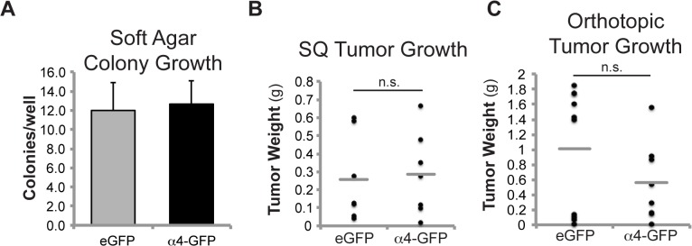
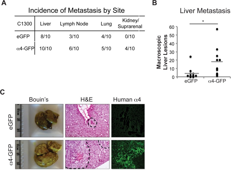
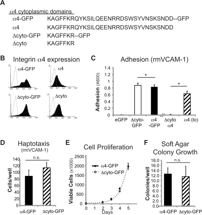
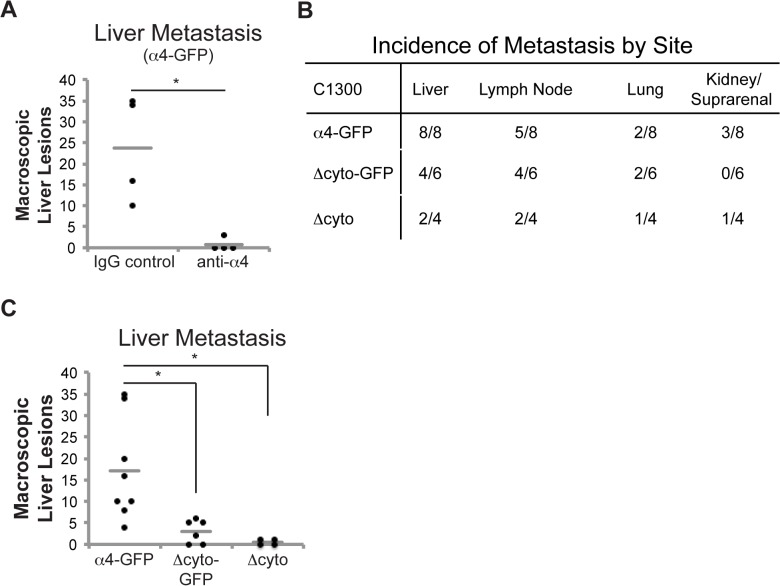
High-risk neuroblastoma is associated with an overall survival rate of 30-50%. Neuroblastoma-expressed cell adhesion receptors of the integrin family impact cell adhesion, migration, proliferation and survival. Integrin α4 is essential for neural crest cell motility during development, is highly expressed on leukocytes, and is critical for transendothelial migration. Thus, cancer cells that express this receptor may exhibit increased metastatic potential. We show that α4 expression in human and murine neuroblastoma cell lines selectively enhances in vitro interaction with the alternatively spliced connecting segment 1 of fibronectin, as well as vascular cell adhesion molecule-1 and increases migration. Integrin α4 expression enhanced experimental metastasis in a syngeneic tumor model, reconstituting a pattern of organ involvement similar to that seen in patients. Accordingly, antagonism of integrin α4 blocked metastasis, suggesting adhesive function of the integrin is required. However, adhesive function was not sufficient, as mutants of integrin α4 that conserved the matrix-adhesive and promigratory function in vitro were compromised in their metastatic capacity in vivo. Clinically, integrin α4 is more frequently expressed in non-MYNC amplified tumors, and is selectively associated with poor prognosis in this subset of disease. These results reveal an unexpected role for integrin α4 in neuroblastoma dissemination and identify α4 as a potential prognostic indicator and therapeutic target.
Conflict of interest statement
Figures
Similar articles
-
p19-INK4d inhibits neuroblastoma cell growth, induces differentiation and is hypermethylated and downregulated in MYCN-amplified neuroblastomas.Hum Mol Genet. 2014 Dec 20;23(25):6826-37. doi: 10.1093/hmg/ddu406. Epub 2014 Aug 7. Hum Mol Genet. 2014. PMID: 25104850
-
ID2 expression in neuroblastoma does not correlate to MYCN levels and lacks prognostic value.Oncogene. 2003 Jan 23;22(3):456-60. doi: 10.1038/sj.onc.1206148. Oncogene. 2003. PMID: 12545167
-
Hox-C9 activates the intrinsic pathway of apoptosis and is associated with spontaneous regression in neuroblastoma.Cell Death Dis. 2013 Apr 11;4(4):e586. doi: 10.1038/cddis.2013.84. Cell Death Dis. 2013. PMID: 23579273 Free PMC article.
-
MYCN in neuronal tumours.Cancer Lett. 2004 Feb 20;204(2):179-87. doi: 10.1016/S0304-3835(03)00454-3. Cancer Lett. 2004. PMID: 15013217 Review.
-
The MYCN oncogene and differentiation in neuroblastoma.Semin Cancer Biol. 2011 Oct;21(4):256-66. doi: 10.1016/j.semcancer.2011.08.001. Epub 2011 Aug 9. Semin Cancer Biol. 2011. PMID: 21849159 Review.
Cited by
-
Role of Corneal Stromal Cells on Epithelial Cell Function during Wound Healing.Int J Mol Sci. 2018 Feb 4;19(2):464. doi: 10.3390/ijms19020464. Int J Mol Sci. 2018. PMID: 29401709 Free PMC article.
-
Status of integrin subunit alpha 4 promoter DNA methylation in colorectal cancer and other malignant tumors: a systematic review and meta-analysis.Res Pharm Sci. 2023 Mar 10;18(3):231-243. doi: 10.4103/1735-5362.371580. eCollection 2023 May-Jun. Res Pharm Sci. 2023. PMID: 37593168 Free PMC article. Review.
-
Novel Aryl Hydrocarbon Receptor Agonist Suppresses Migration and Invasion of Breast Cancer Cells.PLoS One. 2016 Dec 1;11(12):e0167650. doi: 10.1371/journal.pone.0167650. eCollection 2016. PLoS One. 2016. PMID: 27907195 Free PMC article.
-
The silencing effect of miR-30a on ITGA4 gene expression in vitro: an approach for gene therapy.Res Pharm Sci. 2017 Dec;12(6):456-464. doi: 10.4103/1735-5362.217426. Res Pharm Sci. 2017. PMID: 29204174 Free PMC article.
-
A Review of Talin- and Integrin-Dependent Molecular Mechanisms in Cancer Invasion and Metastasis.Int J Mol Sci. 2025 Feb 20;26(5):1798. doi: 10.3390/ijms26051798. Int J Mol Sci. 2025. PMID: 40076426 Free PMC article. Review.
References
-
- Ishola TA, Chung DH (2007) Neuroblastoma. Surg Oncol 16: 149–156. - PubMed
-
- DuBois SG, Kalika Y, Lukens JN, Brodeur GM, Seeger RC, Atkinson JB, et al. (1999) Metastatic sites in stage IV and IVS neuroblastoma correlate with age, tumor biology, and survival. J Pediatr Hematol Oncol 21: 181–189. - PubMed
-
- Ara T, DeClerck YA (2006) Mechanisms of invasion and metastasis in human neuroblastoma. Cancer Metastasis Rev 25: 645–657. - PubMed
-
- Lode HN, Xiang R, Varki NM, Dolman CS, Gillies SD, Reisfeld RA (1997) Targeted interleukin-2 therapy for spontaneous neuroblastoma metastases to bone marrow. J Natl Cancer Inst 89: 1586–1594. - PubMed
-
- Brodeur G, Maris J (2002) Principles and Practice of Pediatric Oncology. In: Pizzo P, Poplack D, editors. Philadelphia, PA: Lippincott Williams & Williams.
Publication types
MeSH terms
Substances
Grants and funding
LinkOut - more resources
Full Text Sources
Other Literature Sources
Medical
