

A Split-and-Merge-Based Uterine Fibroid Ultrasound Image Segmentation Method in HIFU Therapy
- PMID: 25973906
- PMCID: PMC4431844
- DOI: 10.1371/journal.pone.0125738
A Split-and-Merge-Based Uterine Fibroid Ultrasound Image Segmentation Method in HIFU Therapy
Abstract
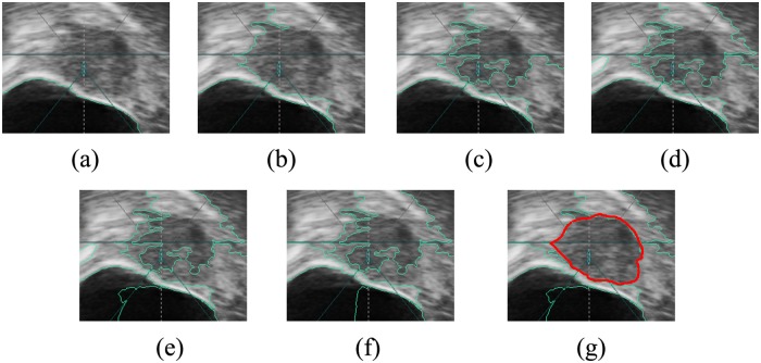
High-intensity focused ultrasound (HIFU) therapy has been used to treat uterine fibroids widely and successfully. Uterine fibroid segmentation plays an important role in positioning the target region for HIFU therapy. Presently, it is completed by physicians manually, reducing the efficiency of therapy. Thus, computer-aided segmentation of uterine fibroids benefits the improvement of therapy efficiency. Recently, most computer-aided ultrasound segmentation methods have been based on the framework of contour evolution, such as snakes and level sets. These methods can achieve good performance, although they need an initial contour that influences segmentation results. It is difficult to obtain the initial contour automatically; thus, the initial contour is always obtained manually in many segmentation methods. A split-and-merge-based uterine fibroid segmentation method, which needs no initial contour to ensure less manual intervention, is proposed in this paper. The method first splits the image into many small homogeneous regions called superpixels. A new feature representation method based on texture histogram is employed to characterize each superpixel. Next, the superpixels are merged according to their similarities, which are measured by integrating their Quadratic-Chi texture histogram distances with their space adjacency. Multi-way Ncut is used as the merging criterion, and an adaptive scheme is incorporated to decrease manual intervention further. The method is implemented using Matlab on a personal computer (PC) platform with Intel Pentium Dual-Core CPU E5700. The method is validated on forty-two ultrasound images acquired from HIFU therapy. The average running time is 9.54 s. Statistical results showed that SI reaches a value as high as 87.58%, and normHD is 5.18% on average. It has been demonstrated that the proposed method is appropriate for segmentation of uterine fibroids in HIFU pre-treatment imaging and planning.
Conflict of interest statement
Figures


Similar articles
-
A superpixel based self-attention network for uterine fibroid segmentation in high intensity focused ultrasound guidance images.Sci Rep. 2025 Jul 1;15(1):21970. doi: 10.1038/s41598-025-08711-x. Sci Rep. 2025. PMID: 40595279 Free PMC article.
-
Multi-scale and shape constrained localized region-based active contour segmentation of uterine fibroid ultrasound images in HIFU therapy.PLoS One. 2014 Jul 25;9(7):e103334. doi: 10.1371/journal.pone.0103334. eCollection 2014. PLoS One. 2014. PMID: 25061939 Free PMC article.
-
A region-based segmentation method for ultrasound images in HIFU therapy.Med Phys. 2016 Jun;43(6):2975-2989. doi: 10.1118/1.4950706. Med Phys. 2016. PMID: 27277046
-
Magnetic resonance-high intensity focused ultrasound (MR-HIFU) therapy of symptomatic uterine fibroids with unrestrictive treatment protocols: A systematic review and meta-analysis.Eur J Radiol. 2019 Nov;120:108700. doi: 10.1016/j.ejrad.2019.108700. Epub 2019 Oct 15. Eur J Radiol. 2019. PMID: 31634683
-
High Intensity Focused Ultrasound Treatment for Uterine Fibroid in a Nigerian Hospital: A Case Report and Review of Literature.West Afr J Med. 2022 Feb 28;39(2):204-207. West Afr J Med. 2022. PMID: 35279044 Review.
Cited by
-
Full coverage path planning algorithm for MRgFUS therapy.Int J Med Robot. 2022 Jun;18(3):e2389. doi: 10.1002/rcs.2389. Epub 2022 Mar 13. Int J Med Robot. 2022. PMID: 35257476 Free PMC article.
-
A superpixel based self-attention network for uterine fibroid segmentation in high intensity focused ultrasound guidance images.Sci Rep. 2025 Jul 1;15(1):21970. doi: 10.1038/s41598-025-08711-x. Sci Rep. 2025. PMID: 40595279 Free PMC article.
-
3D segmentation of uterine fibroids based on deep supervision and an attention gate.Front Oncol. 2025 Mar 13;15:1522399. doi: 10.3389/fonc.2025.1522399. eCollection 2025. Front Oncol. 2025. PMID: 40182051 Free PMC article.
-
Effect of high-intensity focused ultrasound ablation on endometriosis of the abdominal wall.Int J Clin Exp Pathol. 2018 Apr 1;11(4):2118-2124. eCollection 2018. Int J Clin Exp Pathol. 2018. PMID: 31938321 Free PMC article.
References
-
- Stewart EA. Uterine fibroids. Lancet. 2010; 357: 293–8. - PubMed
-
- Kennedy JE. High-intensity focused ultrasound in the treatment of solid tumours. Nat Rev Cancer. 2005; 5: 321–7. - PubMed
-
- Ren XL, Zhou XD, Zhang J, He GB, Han ZH, Zheng MJ, et al. Extracorporeal ablation of uterine fibroids with high-intensity focused ultrasound: imaging and histopathologic evaluation. J Ultrasound Med. 2007; 26: 201–12. - PubMed
Publication types
MeSH terms
LinkOut - more resources
Full Text Sources
Other Literature Sources
Medical
