. 2015 May 14;11(5):e1004836.
doi: 10.1371/journal.ppat.1004836. eCollection 2015 May.

Circulating Pneumolysin Is a Potent Inducer of Cardiac Injury during Pneumococcal Infection

Affiliations

Circulating Pneumolysin Is a Potent Inducer of Cardiac Injury during Pneumococcal Infection

Yasir Alhamdi et al. PLoS Pathog. .

Abstract

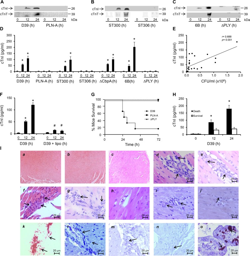
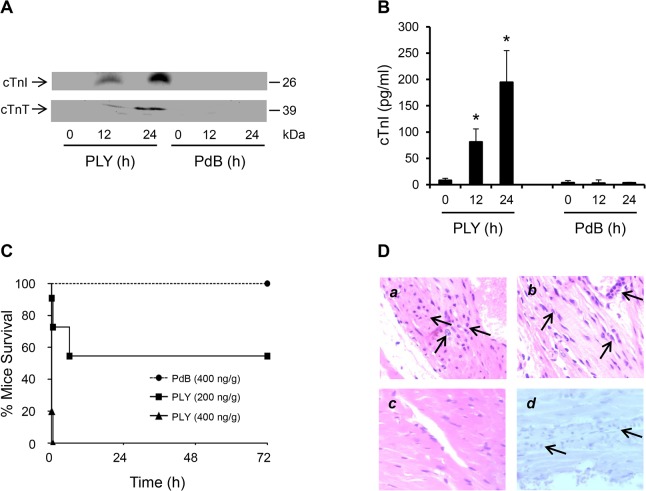
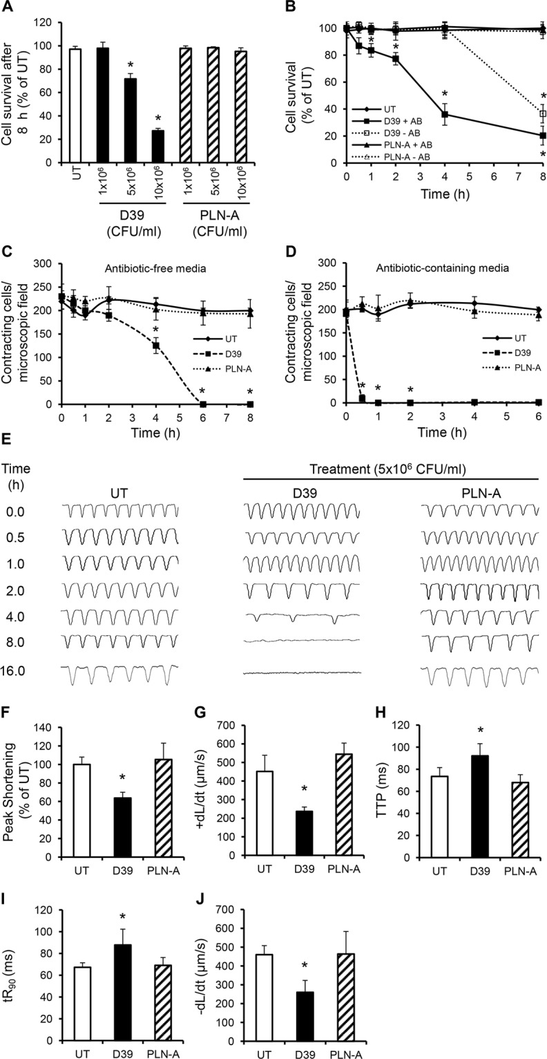
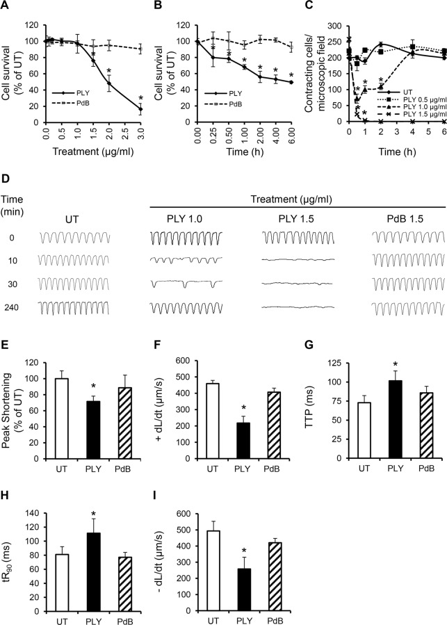
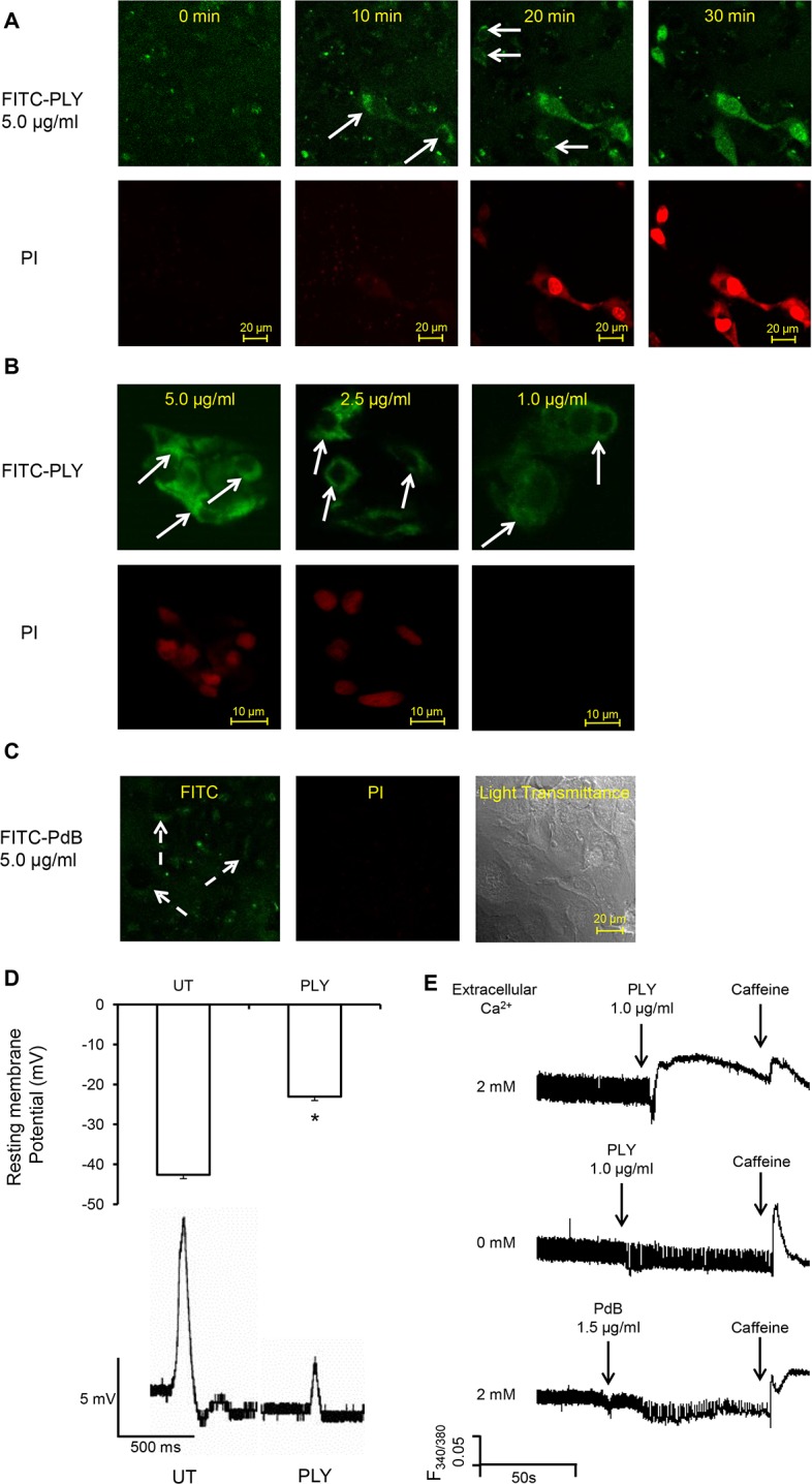
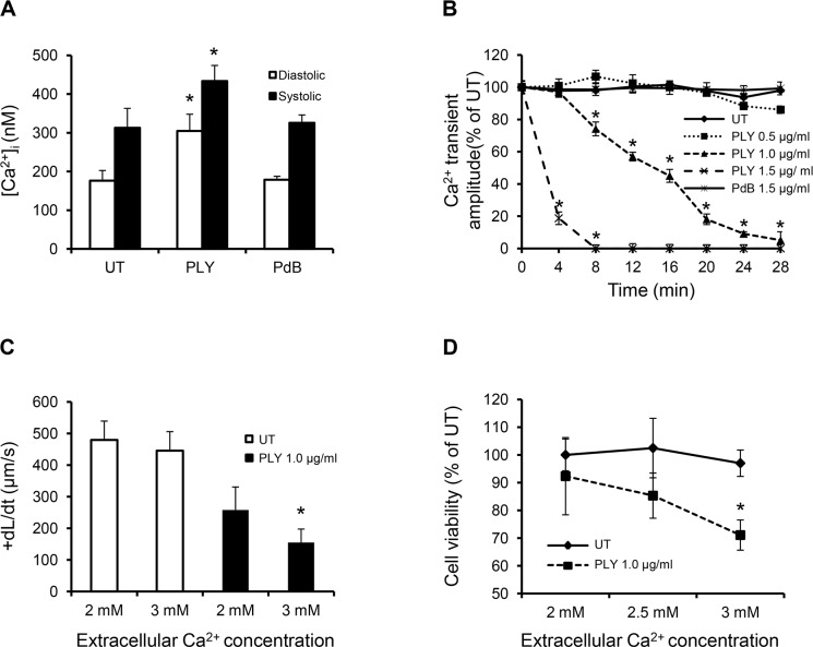
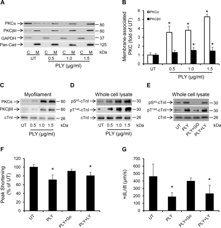
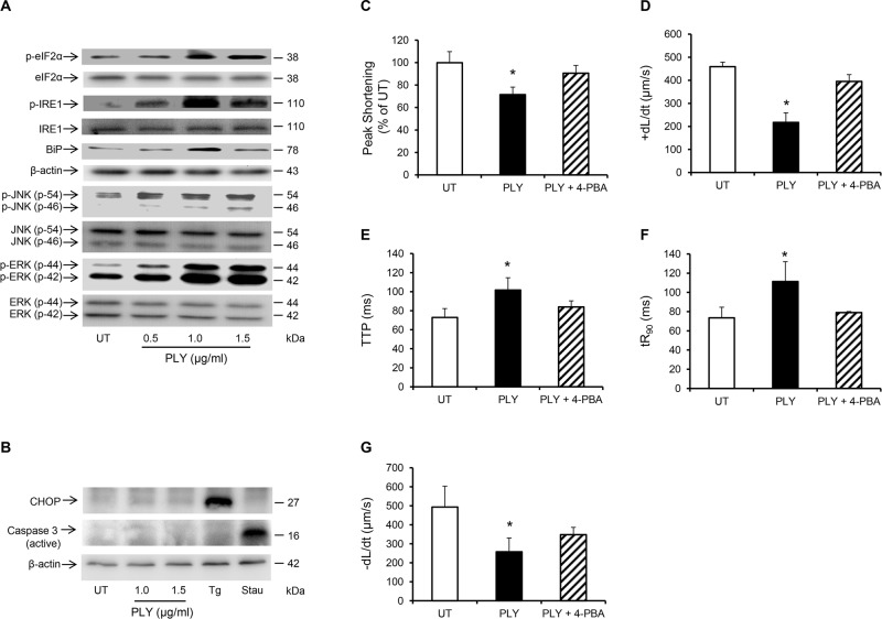
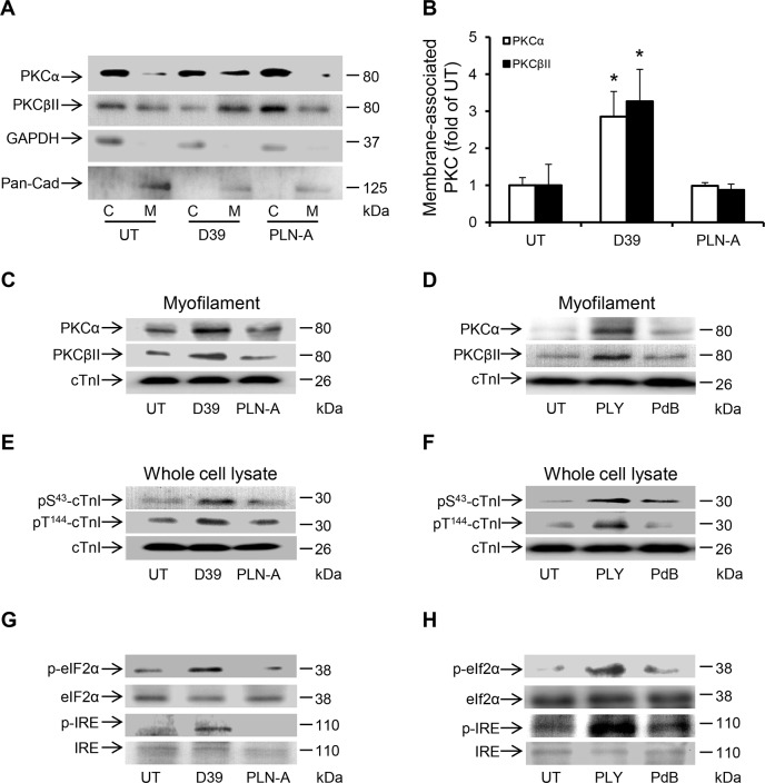
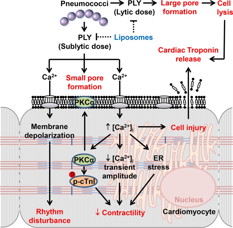
Streptococcus pneumoniae accounts for more deaths worldwide than any other single pathogen through diverse disease manifestations including pneumonia, sepsis and meningitis. Life-threatening acute cardiac complications are more common in pneumococcal infection compared to other bacterial infections. Distinctively, these arise despite effective antibiotic therapy. Here, we describe a novel mechanism of myocardial injury, which is triggered and sustained by circulating pneumolysin (PLY). Using a mouse model of invasive pneumococcal disease (IPD), we demonstrate that wild type PLY-expressing pneumococci but not PLY-deficient mutants induced elevation of circulating cardiac troponins (cTns), well-recognized biomarkers of cardiac injury. Furthermore, elevated cTn levels linearly correlated with pneumococcal blood counts (r=0.688, p=0.001) and levels were significantly higher in non-surviving than in surviving mice. These cTn levels were significantly reduced by administration of PLY-sequestering liposomes. Intravenous injection of purified PLY, but not a non-pore forming mutant (PdB), induced substantial increase in cardiac troponins to suggest that the pore-forming activity of circulating PLY is essential for myocardial injury in vivo. Purified PLY and PLY-expressing pneumococci also caused myocardial inflammatory changes but apoptosis was not detected. Exposure of cultured cardiomyocytes to PLY-expressing pneumococci caused dose-dependent cardiomyocyte contractile dysfunction and death, which was exacerbated by further PLY release following antibiotic treatment. We found that high PLY doses induced extensive cardiomyocyte lysis, but more interestingly, sub-lytic PLY concentrations triggered profound calcium influx and overload with subsequent membrane depolarization and progressive reduction in intracellular calcium transient amplitude, a key determinant of contractile force. This was coupled to activation of signalling pathways commonly associated with cardiac dysfunction in clinical and experimental sepsis and ultimately resulted in depressed cardiomyocyte contractile performance along with rhythm disturbance. Our study proposes a detailed molecular mechanism of pneumococcal toxin-induced cardiac injury and highlights the major translational potential of targeting circulating PLY to protect against cardiac complications during pneumococcal infections.


Conflict of interest statement

The authors have declared that no competing interests exist.

Figures

Fig 1
Fig 1. PLY-expressing but not PLY-deficient pneumococci induce cardiac injury and inflammation.
(A-C) Representative Western blots showing circulating cTnI and cTnT in murine plasma following i.v. injection of D39/PLN-A (n = 4) (1x10⁶ CFU) (A) serotype-1, sequence type 300 and 306 (n = 5) (1x10⁶ CFU) (B), and serotype 6B and PLY-deletion pneumococci (ΔPLY) (1x10⁶ CFU) (C). (D) Mean±SD of circulating cTnI from 4 mice (each group) that survived at 24 h after treatment with D39, PLN-A, ST300, ST306, ΔCbpA, 6B and ΔPLY (1x10⁶ CFU each). *p<0.05 as compared to 0 h. (E) Linear correlation between pneumococcal CFU counts and circulating cTnI levels at various time points (n = 21). (F) Circulating levels of cTnI in plasma of mice following i.v. injection of D39 (1x10⁶ CFU) with or without liposomes (lipo). (n = 3 each group). *p<0.05 as compared to 0 h, #p<0.05 as compared to D39 group. (G) Survival curves of mice infected (i.v.) with D39,PLN-A and ΔPLY (n = 10 for D39, n = 5 for PLN-A, n = 4 for ΔPLY). (H) Circulating levels of cTnI in plasma of mice that died and those that survived within the first 30 h post-D39 infection (n = 4 each group). *p<0.05 as compared to survival group. (I) Pathological examination of murine heart sections after infection with D39, serotype-1, serotype 6B and ΔCbpA pneumococci. H&E representative images (a-h) of murine heart sections under x4 magnification (a-c) and x60 magnification (d-h). Hearts from mice infected with D39 (30 h post infection, d), serotype-1 (e), 6B (f) and ΔCbpA (g) show inflammatory cell infiltration (arrows) at x60 magnification, these are absent in a normal heart section (h). (i-j) Immunohistochemistry images showing absence of pneumococcal capsule staining in heart from mice infected with D39 (i) and serotype-1 (j), despite presence of inflammatory cell infiltrations (arrows). (k) Fresh pneumococci (D39) (arrows) were stained in parallel, as a positive control for pneumococcal capsule staining. (l-n) Representative immunohistochemistry images showing absence of active caspase-3 staining in heart section from mice infected with D39 (l), serotype-1 (m) and 6B (n) despite presence of inflammatory cell infiltrations (arrows). (o) Gut microvilli of a septic mouse showing positive active caspase-3 signal (arrows), was used as a positive control for active caspase-3 staining.
Fig 2
Fig 2. Circulating pore-forming PLY mediates cardiac injury and inflammation in vivo.
(A) Representative Western blots showing circulating cTnI and cTnT in murine plasma following i.v. injection of PLY (200 ng/g) and PdB (400 ng/g). (B) Mean±SD of circulating cTnI from 4 mice (each group) that survived at 24 h after treatment with PLY and PdB. *p<0.05 as compared to PdB group. (C) Survival curves of mice injected (i.v.) with PLY (400 and 200 ng/g) or PdB (400 ng/g) (n = 8 each group). (D) Pathological examination of murine heart sections after infection with purified PLY (200 ng/g) (a, b and d) and PdB (400 ng/g) (c). H&E representative images (a-c) of murine heart sections under x60 magnification showing inflammatory cell infiltrations (arrows) in mice dying within 6 hours (a) and those surviving for 24 hours (b). (c) PdB injection causes no pathological abnormality in murine myocardium. (d) Absence of active caspase-3 staining in murine myocardium following PLY injection.
Fig 3
Fig 3. Wild type (D39) but not PLY-deficient (PLN-A) pneumococci cause cardiomyocyte dysfunction and death in vitro.
(A) Viability of HL-1 cells was assessed at 8 h after incubation with increasing CFUs of D39 and PLN-A. Viability of untreated cells (UT) was set at 100%. Data are presented as Mean±SD (n = 4). (B) Time course of HL-1 cell viability after incubation with 5x10⁶ CFU/ml D39 or PLN-A in the presence or absence of antibiotics (AB). Data are presented as Mean±SD (n = 4). (C) and (D) Effects of D39 and PLN-A (5x10⁶ CFU/ml) on the total number of spontaneously contracting HL-1 cells over time using antibiotic-free (C) and antibiotic-containing media (D). Data are presented as Mean±SD (n = 4). (E) Effects of D39 and PLN-A (5x10⁶ CFU/ml) on the rhythm and rate of contraction of HL-1 cells over time. Typical traces of spontaneous contraction are presented. (F-J) Effects of D39 and PLN-A (5x10⁶ CFU/ml) on Peak Shortening (F), maximum velocity of shortening (+dL/dt) (G), Time to Peak (TTP) (H), Time to 90%-re-lengthening tR90 (I) and maximum velocity of relaxation (-dL/dt) (J) of HL-1 cells after 4 h treatment are presented as Mean±SD (n = 9 from 3 independent experiments). *ANOVA test, p<0.05.
Fig 4
Fig 4. PLY at sub-lytic doses adversely affect cardiomyocyte function in vitro.
(A) Viability of HL-1 cells was assessed 30 min after incubation with increasing concentrations of PLY and PdB using the WST-8 assay. Viability of untreated cells (UT) was set at 100%. Data are presented as Mean±SD. * p<0.05 (n = 4). (B) Time course of HL-1 cell viability after incubation with 1.5 μg/ml PLY or PdB. *p<0.05 (n = 3). (C) Effects of increasing concentrations of PLY on the total number of spontaneously contracting HL-1 cells over time. Data are presented as Mean±SD. *p<0.05 (n = 4). (D) Representative traces of cardiomyocyte contraction before and after PLY and PdB treatment (n = 4). (E-I) Effects of PLY and PdB (1.0 μg/ml) on Peak Shortening (E), +dL/dt (F), TTP (G), tR90 (H) and -dL/dt (I) of HL-1 cells after 30 min treatment are presented as Mean±SD. *p<0.05 (n = 9 from 3 independent experiments).
Fig 5
Fig 5. PLY binds to cardiomyocyte membrane and disrupts Ca2+ homeostasis and membrane potential at sub-lytic concentrations.
(A) HL-1 cells were incubated with 5 μg/ml FITC-PLY and 1.0 μg/ml propidium iodide (PI) to monitor disruption of membrane integrity over time. The localisation of FITC-PLY (green) and PI (red) were recorded using time lapse confocal microscopy (LSM 710, Zeiss) in a maintained environment of 5% CO2 at 37°C. Arrows indicate membrane binding of FITC-PLY. Bar = 20 μm (n = 3). (B) HL-1 cells were treated with 5, 2.5, or 1 μg/ml FITC-PLY along with 1.0 μg/ml PI for 30 min. Following washing in PBS and fixation in 4% PFA, localisation of FITC-PLY and PI were visualized under the same conditions for comparison. Typical images of each dose are presented (n = 3). Arrows indicate membrane binding of FITC-PLY. Bar = 10 μm. (C) HL-1 cells were incubated with 5 μg/ml FITC-PdB and 1.0 μg/ml propidium iodide (PI) to monitor disruption of membrane integrity over time. The localisation of FITC-PdB (green) and PI (red) were recorded as described in (A). Arrows indicate membrane binding of FITC-PdB. Bar = 20 μm (n = 3). (D) Membrane potential changes detected in HL-1 cells following exposure to PLY (1 μg/ml). Top panel: histogram showing typical resting membrane potential under untreated (UT) conditions and following exposure to PLY presented as Mean±SD (n = 3).* p = 0.01. Bottom panel: Typical action potential of HL-1 cells treated without (UT) or with PLY. (E) Changes of [Ca2+]i of HL-1 cardiomyocytes with fura-2am as an indicator were recorded using the IonOptix. Typical traces before and after PLY or PdB treatment in presence and absence of extracellular Ca2+ are presented. Caffeine (10 mM) was used to induce Ca2+ release from sarcoplasmic reticulum stores within cardiomyocytes.
Fig 6
Fig 6. Profound calcium influx induces cardiomyocyte dysfunction and injury in response to sub-lytic PLY.
(A) Mean±SD of both systolic and diastolic [Ca2+]i of HL-cells within 1 min of treatment with PLY/PdB (1.0 μg/ml) from 4 independent experiments are presented. *p<0.05. (B) Change in intracellular Ca2+ transient amplitude over 30 min in response to different concentrations of PLY. Ca2+ transient amplitude at 0 min was set at 100%. Data are presented as Mean±SD (n = 4). (C) and (D) The effects of changing extracellular Ca2+ concentration (in media) of HL-1 cells from 2 to 3 mM on +dL/dt (C) and cell viability (D) in the absence (UT) and presence of 1.0 μg/ml PLY. Mean±SD from 3 independent experiments are presented. * p<0.01 when compared to 2 mM extracellular Ca2+.
Fig 7
Fig 7. PLY activates the PKCα-cTnI axis in HL-1 cardiomyocytes to depress contractility.
(A) Representative Western blots and (B) a band-quantification histogram showing the distribution of PKCα and PKCβII in the cytosol “C” and membrane “M” fractions of HL-1 cells before (UT) and 30 min after PLY-treatment. GAPDH and Pan-Cadherin were used as markers for “C” and “M” fractions, respectively. Data are presented as Mean±SD (n = 3). * p<0.05 compared to UT. (C-E) Typical Western blots showing PLY effects on the association of PKCα and PKCβII with the myofilament fraction (C), the phosphorylation of cTnI at the S43 and T144 PKC-dependent phosphorylation sites (D) and the effects of PKCα inhibitor Go6976 (5 nM) and the PKCβII inhibitor LY333531 (10 nM) on PLY (1.0 μg/ml)-induced phosphorylation of cTnI at S43 and T144 phosphorylation sites (E) in HL-1 cells after 30 min of PLY treatment. cTnI was used as endogenous control (n = 3). (F) and (G) Effects of PLY (1.0 μg/ml) ± Go6976 (5 nM) or LY333531 (10 nM) on Peak Shortening (F) and +dL/dt (G) after 30 min of PLY treatment. Data are presented as Mean±SD (n = 9). *p<0.05 compared to UT.
Fig 8
Fig 8. PLY induces endoplasmic reticulum (ER) stress pathway without progressing to apoptosis in HL-1 cardiomyocytes.
(A) Typical Western blots showing the activation of ER stress markers, (p-elF2α, p-IRE, BiP), JNK and ERK in HL-1 cells 30 min after PLY treatment. (B) Representative western blots showing the effect of PLY on the induction of apoptotic markers CHOP and active caspase-3 after 8 hour of treatment. Thapsigargin (Tg 5 μM) and Staurosporin (Stau 10 μM) were used as positive inducers of CHOP and active caspase-3 respectively. (C-G) Effects of PLY (1.0 μg/ml) and PLY+ 4-Phenylbutyric acid (4-PBA, an ER stress inhibitor, 10 mM) on Peak Shortening (C), (+dL/dt) (D), TTP (E), tR90 (F) and (-dL/dt) (G) of HL-1 cells after 30 min treatment are presented as Mean±SD (n = 9 from 3 independent experiments). * p<0.05.
Fig 9
Fig 9. Activation of PKCα-cTnI pathway and ER stress in murine cardiomyocytes exposed to D39 or PLY.
(A) Representative Western blots and (B) a band-quantification histogram showing the distribution of PKCα and PKCβII in the cytosol “C” and membrane “M” fractions of murine cardiomyocytes under untreated (UT), D39- and PLN-A-infection (1x10⁶ CFU) conditions (24 h post-infection). GAPDH and Pan-Cadherin were used as markers for “C” and “M” fractions, respectively. Data are presented as Mean±SD (n = 3). * p<0.05. (C) and (D) Typical Western blots showing the effects of D39/PLN-A (1x10⁶ CFU) infection (C) and PLY/PdB (200 ng/g) i.v. injection (D) on the association of PKCα and PKCβII with the myofilament fraction of murine cardiomyocytes 24 h post injection. cTnI was used as an endogenous control (n = 3). (E) and (F) Typical Western blots illustrating the phosphorylation of cTnI at the S43 and T144 phosphorylation sites in murine cardiomyocytes following D39/PLN-A infection (E) and PLY/PdB injection (F) (24 h post injection). cTnI was used as an endogenous control (n = 3). (G) and (H) Typical Western blots showing activation of ER stress markers in murine cardiomyocytes at 24 h post-infection with D39/PLN-A (G) and PLY/PdB (H) (n = 3).
Fig 10
Fig 10. The mechanisms and effects of PLY on cardiomyocytes.
High lytic concentrations of PLY induce large pore formation to lyse cells. However, sub-lytic concentrations of PLY bind cellular membrane to induce smaller pores thereby triggering profound Ca2+ influx into cardiomyocytes. The resulting abnormal increment in intracellular Ca2+ concentration [Ca2+]i causes significant membrane depolarization, activation of detrimental signalling pathways (e.g. PKCα-cTnI axis, ER stress) and reductions in the Ca2+ transient amplitude to cause rhythm disturbance and depression in contractile force. Ultimately, Ca2+ overload causes cellular injury which may account for cardiac troponin leakage from cardiomyocytes into the circulation. Toxin-sequestering liposomes offer a potential novel therapeutic intervention against the toxic effects of circulating PLY.

Similar articles

Cited by

References

    1. Ortqvist A, Hedlund J, Kalin M (2005) Streptococcus pneumoniae: epidemiology, risk factors, and clinical features. Semin Respir Crit Care Med 26: 563–574. - PubMed
    1. Viasus D, Garcia-Vidal C, Manresa F, Dorca J, Gudiol F, et al. (2013) Risk stratification and prognosis of acute cardiac events in hospitalized adults with community-acquired pneumonia. J Infect 66: 27–33. 10.1016/j.jinf.2012.09.003 - DOI - PubMed
    1. Corrales-Medina VF, Suh KN, Rose G, Chirinos JA, Doucette S, et al. (2011) Cardiac complications in patients with community-acquired pneumonia: a systematic review and meta-analysis of observational studies. PLoS Med 8: e1001048 10.1371/journal.pmed.1001048 - DOI - PMC - PubMed
    1. Ramirez J, Aliberti S, Mirsaeidi M, Peyrani P, Filardo G, et al. (2008) Acute myocardial infarction in hospitalized patients with community-acquired pneumonia. Clin Infect Dis 47: 182–187. 10.1086/589246 - DOI - PubMed
    1. Corrales-Medina VF, Musher DM, Wells GA, Chirinos JA, Chen L, et al. (2012) Cardiac complications in patients with community-acquired pneumonia: incidence, timing, risk factors, and association with short-term mortality. Circulation 125: 773–781. 10.1161/CIRCULATIONAHA.111.040766 - DOI - PubMed

Publication types

MeSH terms