Review
. 2015 May 13;17(5):577-91.
doi: 10.1016/j.chom.2015.04.008.

An integrative view of microbiome-host interactions in inflammatory bowel diseases

Affiliations
Review

An integrative view of microbiome-host interactions in inflammatory bowel diseases

Marta Wlodarska et al. Cell Host Microbe. .

Abstract

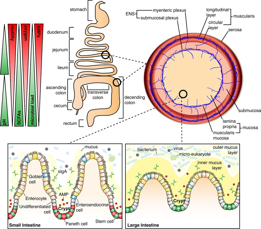
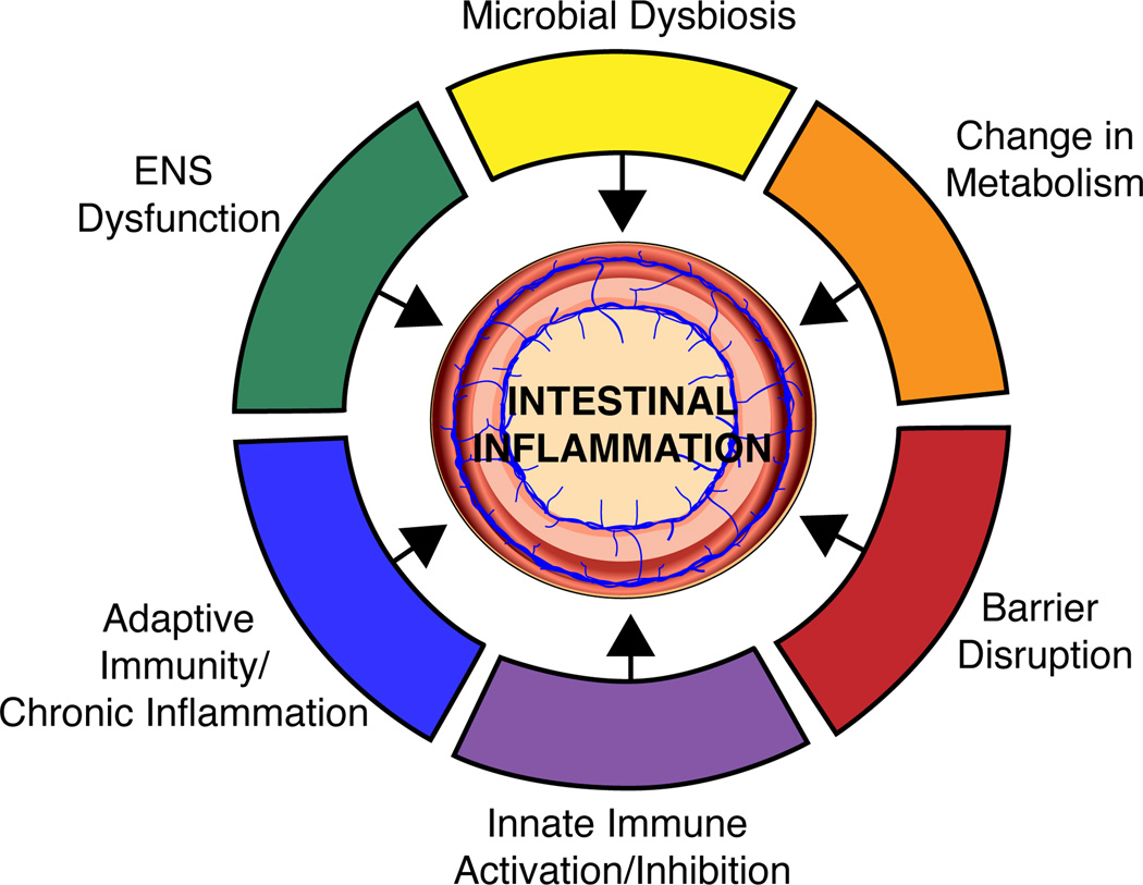
The intestinal microbiota, which is composed of bacteria, viruses, and micro-eukaryotes, acts as an accessory organ system with distinct functions along the intestinal tract that are critical for health. This review focuses on how the microbiota drives intestinal disease through alterations in microbial community architecture, disruption of the mucosal barrier, modulation of innate and adaptive immunity, and dysfunction of the enteric nervous system. Inflammatory bowel disease is used as a model system to understand these microbial-driven pathologies, but the knowledge gained in this space is extended to less-well-studied intestinal diseases that may also have an important microbial component, including environmental enteropathy and chronic colitis-associated colorectal cancer.


Figures

Figure 1
Figure 1. Differentiating features of the small and large intestinal landscape
The small intestine begins after the stomach and is composed of the duodenum, jejunum, and ileum. The ileum joins to the large intestine via the cecum. The large intestine is composed of the ascending colon, transverse colon, descending colon, and rectum. The small intestine has higher oxygen levels and antimicrobial peptide (AMP) production, and increased intestinal motility, whereas in the large intestine, the microbial load is the highest and short-chain fatty acids (SCFAs) are abundant. The entire length of the intestine is lined by a single layer of epithelial cells. Below these cells is the lamina propria (LP), composed of connective tissue that provides the blood supply, lymphatic system, and enervation by the submucosal plexus, which are critical to the function of the intestine. Importantly, the LP houses many immune cells of both the innate and adaptive immune system (not shown). Further enteric enervation occurs in the thin layer of smooth muscle, the muscularis mucosa, which separates the LP from the underlying submucosa. Below the submucosa is a thick muscle layer, the muscularis, composed of an inner circular layer and outer longitudinal layer. Between the two muscle layers is the myenteric plexus, an important component of the enteric nervous system (ENS), which functions to coordinate intestinal peristalsis. The outermost covering of the intestine is the serosa. At the mucosal level the small intestine has long “finger-like” villi that project into the lumen, and which are absent in the large intestine. In the small intestine the crypts contain stem cells, AMP-producing Paneth cells, and undifferentiated cells; the villi contain the differentiated enterocytes, enteroendocrine cells, and goblet cells. In the small intestine goblet cells secrete mucus into the lumen, which has a loose, non-adherent, consistency. In the large intestine, the crypts lack Paneth cells and only contain stem cells and undifferentiated cells; the differentiated cells include enterocytes, enteroendocrine cells, and goblet cells. Here, enterocytes are involved in the production of AMPs and goblet cells secrete mucus that forms a bilayer structure: the inner and outer mucus layers. Although many of the cell types are shared between the small and large intestine, the function of these cells varies depending on the intestinal location.
Figure 2
Figure 2. Intestinal vulnerabilities contributing to IBD
Reduced alpha diversity and a change in the community architecture of the intestinal microbiota is a key phenotype of IBD and may contribute to disease initiation. The composition of the intestinal microbiota heavily influences the metabolic environment of the intestine and specific metabolites have been associated with inflammation. Further microbes influence the activity of both the innate and adaptive arms of the immune system and these interactions can initiate disease as well as maintain chronic inflammation. Intestinal microbes have been shown to produce a variety of neurotransmitters and are also critical for ENS development and function; these microbial-induced modifications to the ENS can translate to downstream effects on the mucosal immune system.

References

    1. Al-Saleem T, Al-Mondhiry H. Immunoproliferative small intestinal disease (IPSID): a model for mature B-cell neoplasms. Blood. 2005;105:2274–2280. - PubMed
    1. Albenberg L, Esipova TV, Judge CP, Bittinger K, Chen J, Laughlin A, Grunberg S, Baldassano RN, Lewis JD, Li H, et al. Correlation between intraluminal oxygen gradient and radial partitioning of intestinal microbiota. Gastroenterology. 2014;147:1055–1063.e8. - PMC - PubMed
    1. Alfellani MA, Stensvold CR, Vidal-Lapiedra A, Onuoha ESU, Fagbenro-Beyioku AF, Clark CG. Variable geographic distribution of Blastocystis subtypes and its potential implications. Acta Trop. 2013;126:11–18. - PubMed
    1. An D, Oh SF, Olszak T, Neves JF, Avci FY, Erturk-Hasdemir D, Lu X, Zeissig S, Blumberg RS, Kasper DL. Sphingolipids from a symbiotic microbe regulate homeostasis of host intestinal natural killer T cells. Cell. 2014;156:123–133. - PMC - PubMed
    1. Arpaia N, Campbell C, Fan X, Dikiy S, van der Veeken J, deRoos P, Liu H, Cross JR, Pfeffer K, Coffer PJ, et al. Metabolites produced by commensal bacteria promote peripheral regulatory T-cell generation. Nature. 2013;504:451–455. - PMC - PubMed

Publication types

MeSH terms