Review
. 2015 Oct;237(2):135-45.
doi: 10.1002/path.4563. Epub 2015 Jun 15.

Microenvironmental control of stem cell fate in intestinal homeostasis and disease

Affiliations
Review

Microenvironmental control of stem cell fate in intestinal homeostasis and disease

Sujata Biswas et al. J Pathol. 2015 Oct.

Abstract

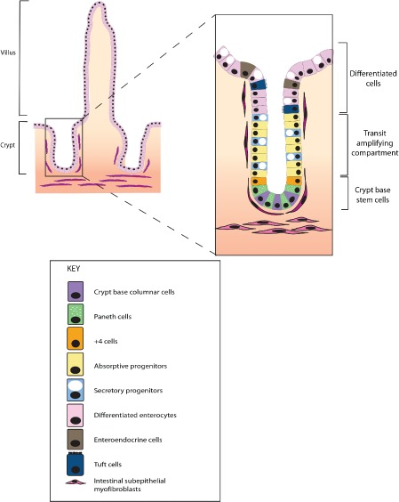
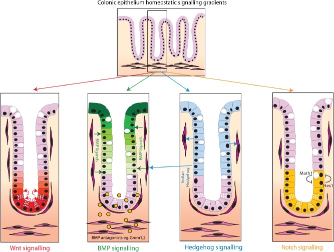
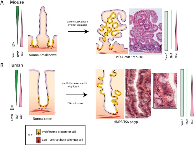
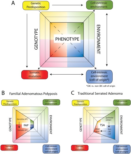
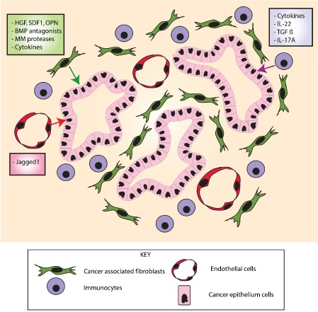
The conventional model of intestinal epithelial architecture describes a unidirectional tissue organizational hierarchy with stem cells situated at the crypt base and daughter cells proliferating and terminally differentiating as they progress along the vertical (crypt-luminal) axis. In this model, the fate of a cell that has left the niche is determined and its lifespan limited. Evidence is accumulating to suggest that stem cell control and daughter cell fate determination is not solely an intrinsic, cell autonomous property but is heavily influenced by the microenvironment including paracrine, mesenchymal, and endogenous epithelial morphogen gradients. Recent research suggests that in intestinal homeostasis, stem cells transit reversibly between states of variable competence in the niche. Furthermore, selective pressures that disrupt the homeostatic balance, such as intestinal inflammation or morphogen dysregulation, can cause committed progenitor cells and even some differentiated cells to regain stem cell properties. Importantly, it has been recently shown that this disruption of cell fate determination can lead to somatic mutation and neoplastic transformation of cells situated outside the crypt base stem cell niche. This paper reviews the exciting developments in the study of stem cell dynamics in homeostasis, intestinal regeneration, and carcinogenesis, and explores the implications for human disease and cancer therapies.

Keywords: chronic inflammation; colon; neoplasia.


Figures

Figure 1
Figure 1
Intestinal crypt architecture and cell types. The intestinal crypt is the basic functional unit of the gut. In the small intestine, several crypts contribute to finger‐like projections called villi. In homeostasis, the stem cells (crypt base columnar and +4 cells) are restricted to the crypt base stem cell niche. Immediate stem cell progeny divide rapidly in the bottom half of the crypt, called the transit amplifying zone. Terminal differentiation occurs in the upper part of the crypt, with fully differentiated cells eventually being shed into the intestinal lumen. Under homeostatic conditions in the mammalian gut, transit along the crypt–luminal axis takes 5–7 days.
Figure 2
Figure 2
Key morphogen signalling pathway gradients in intestinal homeostasis. Intestinal homeostasis and cell fate determination are maintained by a complex interaction of polarized epithelial and mesenchymal morphogen signalling pathways. Major pathways include (A) Wnt signalling. Wnt ligands, secreted from the intestinal subepithelial myofibrobasts and the Paneth cells, act predominantly at the base of the crypt (red cells) to maintain stem cell function and transit amplifying cell proliferation; (B) Bone morphogenetic protein signalling. BMP ligands are produced predominantly by the mesenchymal cells, although BMP2 is also expressed by epithelial cells. BMP represses Wnt signalling 26 and promotes cell differentiation and apoptosis, acting predominantly in the upper part of the crypt and villus (green cells). There is reduced BMP activity at the base of the crypt, partly through diffusion gradients of the ligands but also secondary to the restricted expression of ligand‐sequestering BMP antagonists from the sub‐crypt myofibroblasts; (C) Hedgehog signalling. Indian Hedgehog (IHH) is the main ligand and is expressed by epithelial cells in the upper part of the crypt (blue cells). IHH acts upon and maintains the myofibroblasts. This has a secondary effect on the epithelium through promotion of BMP ligand expression; (D) Notch signalling. Notch regulates cell fate through cell‐to‐cell contact in the stem cell and transit amplifying zone at the base of the crypt (yellow cells). Notch activation via Hes1 transcription factor and lateral inhibition via Math1 transcription factor regulate enterocyte or secretory cell fate, respectively.
Figure 3
Figure 3
Compartmental expression switch of GREM1 expression initiates neoplasia from an expanded progenitor cell population. Aberrant epithelial expression of the BMP antagonist GREM1 disrupts the polarized BMP gradient and dysregulates cell fate determination along the crypt–villus axis of the intestine. This results in the expansion of a proliferating progenitor population which forms ectopic crypt foci. (A) Mouse. In the mouse, ectopic crypts are seen on the villi of the small intestine and dysplasia arises from within these intravillus lesions. (B) Human. In human hereditary mixed polyposis syndrome (HMPS) and sporadic traditional serrated adenoma (TSA) lesions, ectopic crypts can be seen developing orthogonally to the axis of the crypt. Coloured bars represent morphogen gradients in the normal and pathological states. Blue squares represent physiological Grem1 expression from pericryptal myofibroblasts. CBC stem cells are red and progenitor cells are yellow.
Figure 4
Figure 4
Genetic and environmental influences variably contribute to cancer phenotype in different colorectal cancer subtypes. A key feature of Darwinian evolutionary theory is that cancer phenotype is determined by a combination of genetic and environmental influences. (A) We propose an update of this important maxim by dividing genotype into genetic predisposition and somatic mutation, and environment into cell‐intrinsic (cell‐of‐origin) and cell‐extrinsic environment. There is considerable interaction between these different influences, eg the cell of origin selects for an optimal somatic mutation (arrows). The variety of cancer phenotypes can be related to the variable importance of these four different influences in tumour development. (B) Familial adenomatous polyposis (FAP). In FAP, germline APC mutation in epithelial stem cells predominantly drives tumourigenesis without the requirement for an altered cell‐intrinsic or cell‐extrinsic microenvironment. (C) Traditional serrated adenoma (TSA). In contrast, in sporadic TSAs, the aberrant morphogen environment provoked by epithelial GREM1 expression leads to cancer initiation from cells outside of the crypt base. This selects for optimal somatic mutations that differ from conventional Wnt‐driven carcinogenic pathways. CBC cell = crypt base columnar cell.
Figure 5
Figure 5
Stromal signalling pathways affecting epithelial cell stem cell function in the cancer microenvironment. Different cancer stromal elements express numerous factors that act upon the epithelium to modulate epithelial cancer stem cell function. The cancer‐associated fibroblasts express BMP antagonists and factors such as hepatocyte growth factor (HGF) and osteopontin (OPN). Tumour‐infiltrating immune cells secrete cytokines such as IL‐22 and IL‐17, and endothelial cells interact with the epithelium via expression of Jagged1, regulating the Notch pathway.

Similar articles

Cited by

References

    1. Cheng H, Leblond CP. Origin, differentiation and renewal of the four main epithelial cell types in the mouse small intestine. I. Columnar cell. Am J Anat 1974; 141 : 461–479. - PubMed
    1. Potten CS. Extreme sensitivity of some intestinal crypt cells to X and gamma irradiation. Nature 1977; 269: 518‐521. - PubMed
    1. Scoville DH, Sato T, He XC, et al. Current view: intestinal stem cells and signaling. Gastroenterology 2008; 134: 849‐864. - PubMed
    1. Medema JP, Vermeulen L. Microenvironmental regulation of stem cells in intestinal homeostasis and cancer. Nature 2011; 474: 318‐326. - PubMed
    1. Ritsma L, Ellenbroek SI, Zomer A, et al. Intestinal crypt homeostasis revealed at single‐stem‐cell level by in vivo live imaging. Nature 2014; 507: 362‐365. - PMC - PubMed

MeSH terms