Review
. 2015 Jun 17;26(6):975-86.
doi: 10.1021/acs.bioconjchem.5b00191. Epub 2015 May 22.

HaloTag technology: a versatile platform for biomedical applications

Affiliations
Review

HaloTag technology: a versatile platform for biomedical applications

Christopher G England et al. Bioconjug Chem. .

Abstract

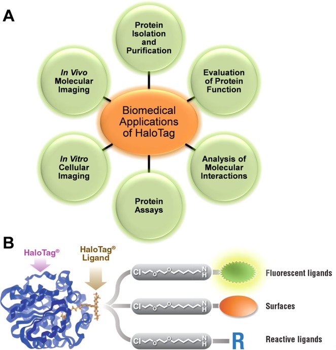
Exploration of protein function and interaction is critical for discovering links among genomics, proteomics, and disease state; yet, the immense complexity of proteomics found in biological systems currently limits our investigational capacity. Although affinity and autofluorescent tags are widely employed for protein analysis, these methods have been met with limited success because they lack specificity and require multiple fusion tags and genetic constructs. As an alternative approach, the innovative HaloTag protein fusion platform allows protein function and interaction to be comprehensively analyzed using a single genetic construct with multiple capabilities. This is accomplished using a simplified process, in which a variable HaloTag ligand binds rapidly to the HaloTag protein (usually linked to the protein of interest) with high affinity and specificity. In this review, we examine all current applications of the HaloTag technology platform for biomedical applications, such as the study of protein isolation and purification, protein function, protein-protein and protein-DNA interactions, biological assays, in vitro cellular imaging, and in vivo molecular imaging. In addition, novel uses of the HaloTag platform are briefly discussed along with potential future applications.


Figures

Figure 1
Figure 1
Applications of the versatile HaloTag platform. (A) The HaloTag protein tagging system is utilized for several applications, including protein isolation and purification, evaluation of protein function, analysis of molecular interactions, protein assays, in vitro cellular imaging, and in vivo molecular imaging. (B) Representation of the HaloTag system, in which the HaloTag protein forms a covalent bond with a specific HaloTag ligand. Each HaloTag ligand contains a binding group and functional moiety, such as fluorescent molecules for intracellular and extracellular purposes, surface ligands for protein immobilization with resins or slides, and reactive ligands for imaging purposes. Reprinted with permission from ref (11). Copyright 2012 Urh and Rosenberg.
Figure 2
Figure 2
Functional human kinases isolated and purified from HEK-293 cells using the HaloTag platform. (A) Five human kinases were selected for isolation and purification. (B) To compare the efficiency of HaloTag to that of other protein tagging systems, PKCγ and PI3Kγ were transiently expressed in HEK-293 cells using four protein labeling protocols, including HaloTag, FLAG, 3× FLAG, and His-Tag. Purified proteins were analyzed by SDS-PAGE. Each protein tagging method resulted in purified protein, yet only HaloTag displayed a single band. The arrow denotes the expected molecular weight of the protein. (C) Protein recovery was determined using normalized volumes of soluble lysate (S), unbound fractions (FT), and purified protein (Y) with the addition of a protease using SDS-PAGE and western blot analysis. (D) The HaloTag platform provided the highest purity of protein for both kinases, as compared to that with the other systems. The percent recovery was also shown to be much higher for the HaloTag system. Reprinted with permission from ref (20). Copyright 2011 Elsevier.
Figure 3
Figure 3
Hydrophobic molecules induce degradation of HaloTag proteins. (A) Chemical structure of six hydrophobic HaloTag ligands. (B) Human embryonic kidney cell line, HEK 293T, stably expressing luciferase-modified HaloTag protein was used to measure the biological activity of hydrophobic HaloTag ligands. (C) NIH-3T3 xenografts expressing HaloTag protein were implanted into mice. Tumor growth was monitored in the presence of a hydrophobic HaloTag ligand (HyT13). Reprinted with permission from ref (49). Copyright 2011 Macmillan Publishers Limited.
Figure 4
Figure 4
Pull-down assays for the discovery of protein complexes. (A) Schematic illustration of HaloTag pull-down assays, in which a single HaloTag construct encoding a bait protein is stably transfected into a cell line. The bait protein interacts with additional proteins, at which time cells are lysed and captured using HaloLink resin. Pure proteins can be eluted using a detergent (e.g., SDS), or protein complexes attached the bait protein can be eluted using TEV cleavage. Reprinted with permission from ref (57). Copyright 2014 JoVE. (B) A pull-down assay was performed using HaloTag-modified RpoA to determine the efficiency of HaloTag to extract multiprotein complexes. M, molecular weight marker; 1, unbound proteins; 2, washed proteins; 3, eluted proteins after TEV cleavage; 4, eluted proteins after removal of TEV; and 5, concentrated protein sample. Arrows indicate recombinant HaloTag in lane 1 and cleaved RpoA in lane 3. In addition, a protein interaction map was constructed from data using MALDI-MS/MS. Reprinted with permission from ref (61). Copyright 2012 Peterson and Kwon.
Figure 5
Figure 5
Intracellular imaging of HaloTag-modified amylase using immunofluorescence microscopy. (A) Two HaloTag–amylase proteins (HaloTag A and HaloTag B) were constructed, as the exact translational start site for salivary amylase has not been identified. (B) Expression of nonconjugated HaloTag (Halo) and both HaloTag A and B proteins was examined. The top band represents the HaloTag complex, whereas the smaller band is indicative of pure HaloTag without amylase attached. (C) Halo, HaloTag A, and HaloTag B were labeled with a HaloTag ligand (TMR-Green), and secretory granules were labeled with an anti-amylase antibody (shown in red). Both HaloTag A and B show colocalization with endogenous amylase, indicating that both were in secretory granules. Scale bar = 10 μm. Reprinted with permission from ref (92). Copyright 2013 the American Physiological Society.
Figure 6
Figure 6
PET imaging of 4T1 tumor-bearing mice with novel HaloTag ligands. (A) Chemical structure of NOTA-HTL2G-S and NOTA-HTL2G-L, with different lengths of PEG. (B) PET imaging of mice with two 4T1 xenografts; the left tumor does not express HaloTag protein, and the right tumor expresses HaloTag protein. Mice were injected with the short (64Cu-NOTA-HTL2G-S) or long (64Cu-NOTA-HTL2G-L) form of the HaloTag ligand. In addition, a blocking agent was used with 64Cu-NOTA-HTL2G-L. Accumulation of 64Cu-NOTA-HTL2G-L ligand can be seen in HaloTag tumors from 3 to 24 h postinjection. Reprinted with permission from ref (120). Copyright 2013 AJTR.

References

    1. Toyama B. H.; Hetzer M. W. (2013) Protein homeostasis: live long, won’t prosper. Nat. Rev. Mol. Cell Biol. 14, 55–61. - PMC - PubMed
    1. Robinson C. V.; Sali A.; Baumeister W. (2007) The molecular sociology of the cell. Nature 450, 973–982. - PubMed
    1. Hanash S. (2003) Disease proteomics. Nature 422, 226–232. - PubMed
    1. Hanash S. M.; Pitteri S. J.; Faca V. M. (2008) Mining the plasma proteome for cancer biomarkers. Nature 452, 571–579. - PubMed
    1. Kolch W.; Mischak H.; Pitt A. R. (2005) The molecular make-up of a tumour: proteomics in cancer research. Clin. Sci. 108, 369–383. - PubMed

Publication types