. 2016 Mar;26(2):155-66.
doi: 10.1111/bpa.12265. Epub 2015 Jun 4.

Pathological and Clinical Spectrum of Progressive Supranuclear Palsy: With Special Reference to Astrocytic Tau Pathology

Affiliations

Pathological and Clinical Spectrum of Progressive Supranuclear Palsy: With Special Reference to Astrocytic Tau Pathology

Yuichi Yokoyama et al. Brain Pathol. 2016 Mar.

Abstract

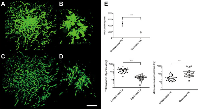
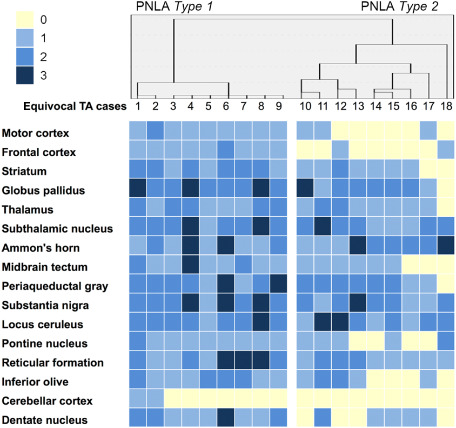
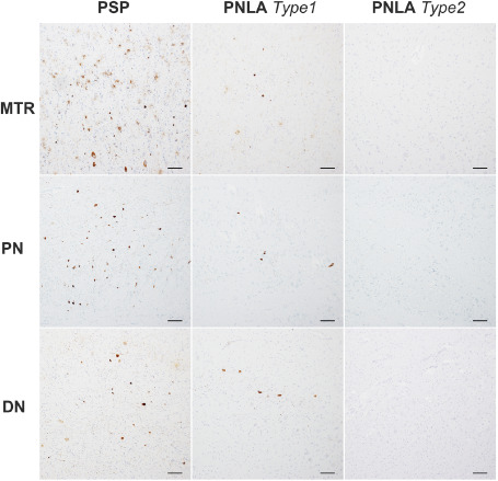
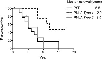
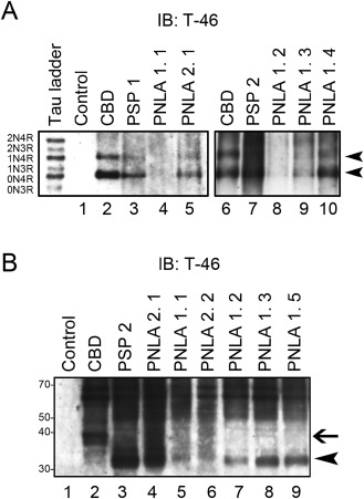
Progressive supranuclear palsy (PSP) is a four-repeat tauopathy with tau-positive, argyrophilic tuft-shaped astrocytes (TAs). We performed a pathological and clinical investigation in 40 consecutive autopsied Japanese patients with pathological diagnoses of PSP or PSP-like disease. Unequivocal TAs were present in 22 cases, all of which were confirmed to be PSP. Such TAs were hardly detected in the other 18 cases, which instead exhibited tau-positive, argyrophilic astrocytes, appearing as comparatively small clusters with central nuclei of irregularly shaped, coarse structures (equivocal TAs). Cluster analysis of the distribution pattern of tau-related pathology for these 18 cases identified two subgroups, pallido-nigro-luysian atrophy (PNLA) Type 1 (n = 9) and Type 2 (n = 9), the former being distinguished from the latter by the presence of tau-related lesions in the motor cortex, pontine nucleus and cerebellar dentate nucleus in addition to the severely affected PNL system. The duration from symptom onset until becoming wheelchair-bound was significantly longer in PNLA Type 1. Immunoblotting of samples from the three disease conditions revealed band patterns of low-molecular-mass tau fragments at ∼35 kDa. These findings shed further light on the wide pathological and clinical spectrum of four-repeat tauopathy, representing PSP in the broad sense rather than classical PSP.

Keywords: glial tau pathology; pallido-nigro-luysian atrophy; progressive supranuclear palsy; tauopathy; tuft-shaped astrocyte.


Conflict of interest statement

The authors have no conflicts of interest to declare.

Figures

Figure 1
Figure 1
Grading scale for tau‐related pathology. The occurrence of tau‐related lesions was assessed using a 4‐point rating scale: 0, absent; 1, sparse; 2, moderate; 3, numerous. Pontine nuclei immunostained with AT8. Bar = 200 μm.
Figure 2
Figure 2
A–D. Tau‐positive, argyrophilic unequivocal tuft‐shaped (uTA) and equivocal tuft‐shaped (eTA) astrocytes. In uTAs, tau appears to have accumulated mainly in the cell processes, showing a radiating, fine, long curvilinear pattern of tau positivity (A) and argyrophilia (C). On the other hand, in eTAs, tau appears to have accumulated mainly in the perikarya, showing a thick and coarse pattern of tau positivity (B) and argyrophilia (D). Note that uTAs (A, C) show larger expanses of tau immunoreactivity and argyrophilia than eTAs (B, D). UTAs (E) and eTAs (F) are also recognizable with another phosphorylation‐dependent anti‐tau antibody. (A, B) AT8 immunostaining; (C, D) Gallyas–Braak staining. (E, F) T212 immunostaining. Bars = 10 μm in A–F.
Figure 3
Figure 3
A–D. Three‐dimensional (3D) reconstruction of the astrocytic tau pathology: a AT8‐positive uTA (A, C) and an AT8‐positive eTA (B, D). 3D images obtained by LSM software (in the ZEN transparent mode) (A, B) and by IMARIS software based on fluorescence intensity for morphometric analysis (C, D). E. Morphometric analysis using IMARIS indicated significant differences in the volumes, the total numbers of particles and the mean volumes per particle between uTAs and eTAs (****P < 0.0001). The values shown as vertical lines indicate the mean with 95% confidence interval (CI). Note that with regard to each parameter, there is no overlap in the 95% CI between uTAs and eTAs.
Figure 4
Figure 4
A–D. Pre‐embedding immunoelectron microscopy with AT8. In an uTA, AT8‐positive products can be seen as radiating slender structures extending from the small perinuclear area (A); in an area of another TA at higher magnification, although indistinct, a bundle of straight tubule‐like structures can be seen (C). In an eTA, AT8‐positive products are evident as thick and coarse structures located mainly in the relatively large cytoplasm (B); higher magnification view of the area indicated by the arrow in B, showing an aggregate of AT8‐positive products; similar straight tubule‐like structures are dimly evident (D). E, F. Conventional electron microscopy shows bundles of tubular structures in the cell process of an uTA (E) and the cell body of an eTA (F). (A–F) Putamen. Bars: (A, B) 5 μm; (C, D) 500 nm; (E, F) 200 nm.
Figure 5
Figure 5
Cluster analysis and distribution of AT8‐positive tau lesions in 18 cases belonging to the eTA group. Dendrogram of cluster analysis of the distribution of AT8‐positive tau lesions (upper panel). Distribution of AT8‐positive tau lesions (lower panel). The severity is also shown in each case and in each region.
Figure 6
Figure 6
Representative densities of AT8‐positive tau lesions seen in the motor cortex (MTR), pontine nuclei (PN) and cerebellar dentate nucleus (DN) in PSP, and PNLAType 1 and Type 2 cases. In these regions, the scores for tau‐related pathology were generally 2–3 in PSP, 1–2 in PNLAType 1 and 0–1 in PNLAType 2. Bars = 100 μm.
Figure 7
Figure 7
Kaplan–Meier survival analysis of the period from symptom onset until the patient becoming wheelchair‐bound. Survival time was significantly longer in PNLAType 1 than in PSP and PNLAType 2 (P < 0.05).
Figure 8
Figure 8
A. Immunoblot analysis of the sarkosyl‐insoluble fraction after dephosphorylation in two PSP (PSP 1, 2: lanes 3, 7) cases, and four PNLAType 1 (PNLA 1. 1–4: lanes 4, 8, 9, 10) and one Type 2 (PNLA 2. 1: lane 5) cases, as well as control (lane 1) and CBD (lanes 2, 6) cases. Sarkosyl‐insoluble tau in cases of PSP and PNLAType 1 and Type 2, as well as CBD consists predominantly of 4R tau isoforms (0N4R and 1N4R, arrowheads). Six recombinant human tau isoforms are indicated on the left (tau ladder). B. Immunoblot analysis of the sarkosyl‐insoluble fraction before dephosphorylation. The pattern of low‐molecular‐mass tau fragments in a PSP (PSP 2: lane 3) case, and 4 PNLAType 1 (PNLA 1. 1–3, 5: lanes 5, 7, 8, 9) and 2 Type 2 (PNLA 2. 1, 2: lanes 4, 6) cases shows a prominent band of 33 kDa (arrowhead), while that in a CBD (lane 2) case shows a prominent band of 37 kDa (arrow).

References

    1. Ahmed Z, Josephs KA, Gonzalez J, DelleDonne A, Dickson DW (2008) Clinical and neuropathologic features of progressive supranuclear palsy with severe pallido‐nigro‐luysial degeneration and axonal dystrophy. Brain 131:460–472. - PubMed
    1. Ahmed Z, Tabrizi SJ, Li A, Houlden H, Sailer A, Lees AJ et al (2010) A Huntington's disease phenocopy characterized by pallido‐nigro‐luysian degeneration with brain iron accumulation and p62‐positive glial inclusions. Neuropathol Appl Neurobiol 36:551–557. - PubMed
    1. Ahmed Z, Bigio EH, Budka H, Dickson DW, Ferrer I, Ghetti B et al (2013) Globular glial tauopathies (GGT): consensus recommendations. Acta Neuropathol 126:537–544. - PMC - PubMed
    1. Arai T, Ikeda K, Akiyama H, Nonaka T, Hasegawa M, Ishiguro K et al (2004) Identification of amino‐terminally cleaved tau fragments that distinguish progressive supranuclear palsy from corticobasal degeneration. Ann Neurol 55:72–79. - PubMed
    1. Arima K (2006) Ultrastructural characteristics of tau filaments in tauopathies: immuno‐electron microscopic demonstration of tau filaments in tauopathies. Neuropathology 26:475–483. - PubMed

Publication types