. 2015 May 14:5:9865.
doi: 10.1038/srep09865.

Tracking of dendritic cell migration into lymph nodes using molecular imaging with sodium iodide symporter and enhanced firefly luciferase genes

Affiliations

Tracking of dendritic cell migration into lymph nodes using molecular imaging with sodium iodide symporter and enhanced firefly luciferase genes

Ho Won Lee et al. Sci Rep. .

Erratum in

Abstract

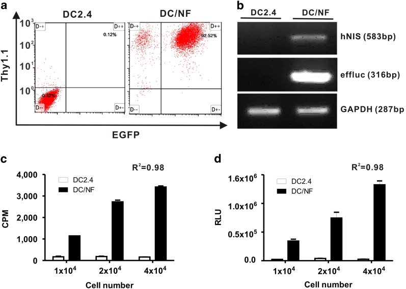
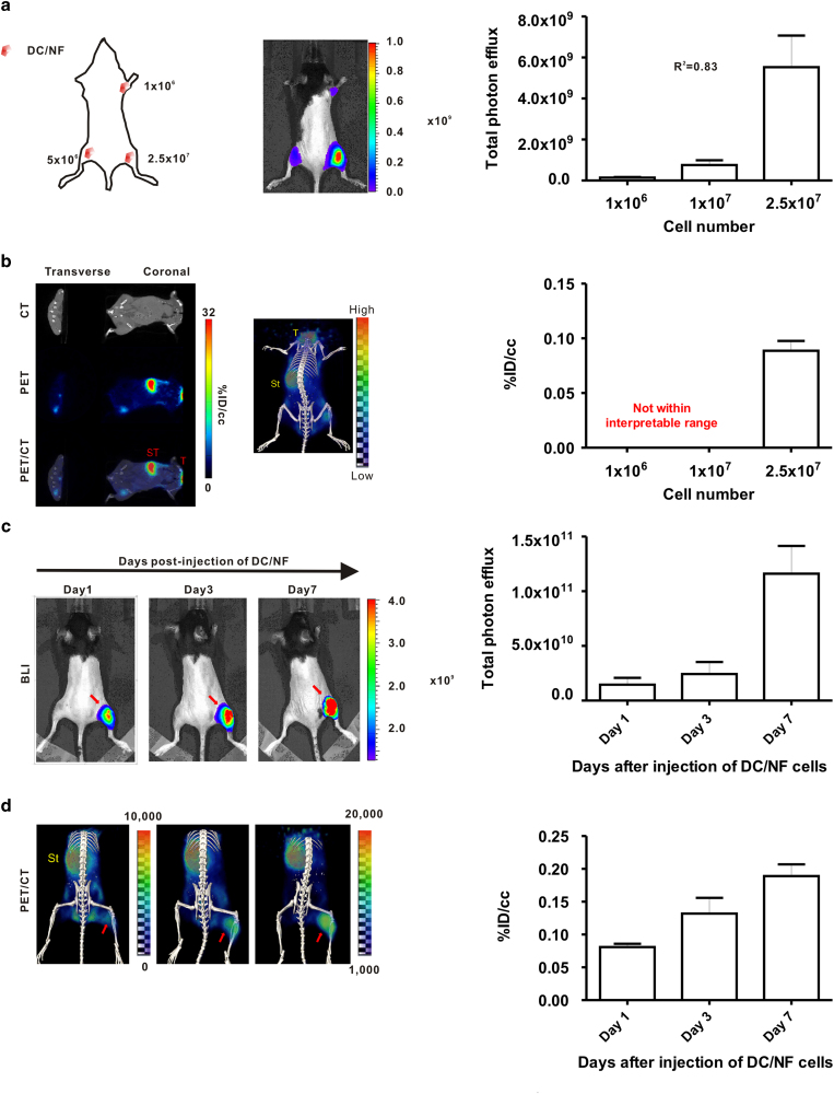
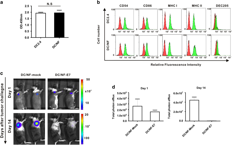
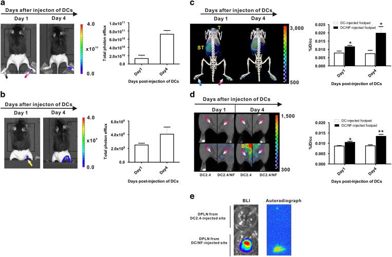
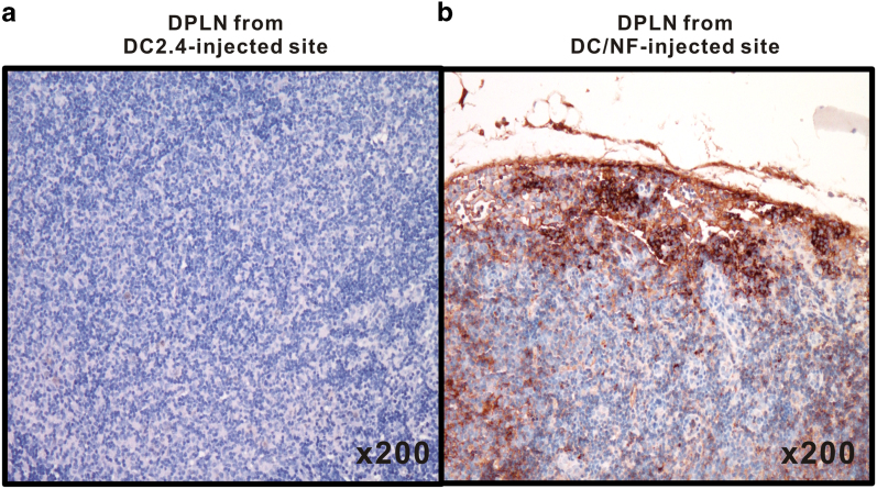
We sought to evaluate the feasibility of molecular imaging using the human sodium iodide symporter (hNIS) gene as a reporter, in addition to the enhanced firefly luciferase (effluc) gene, for tracking dendritic cell (DCs) migration in living mice. A murine dendritic cell line (DC2.4) co-expressing hNIS and effluc genes (DC/NF) was established. For the DC-tracking study, mice received either parental DCs or DC/NF cells in the left or right footpad, respectively, and combined I-124 PET/CT and bioluminescence imaging (BLI) were performed. In vivo PET/CT imaging with I-124 revealed higher activity of the radiotracer in the draining popliteal lymph nodes (DPLN) of the DC/NF injection site at day 1 than DC injection site (p < 0.05). The uptake value further increased at day 4 (p < 0.005). BLI also demonstrated migration of DC/NF cells to the DPLNs at day 1 post-injection, and signals at the DPLNs were much higher at day 4. These data support the feasibility of hNIS reporter gene imaging in the tracking of DC migration to lymphoid organs in living mice. DCs expressing the NIS reporter gene could be a useful tool to optimize various strategies of cell-based immunotherapy.


Figures

Figure 1
Figure 1. Establishment of dendritic cells co-expressing the hNIS and effluc genes.
(a) Flow cytometric analysis of parental DC2.4 and DC/NF cells using an APC-Cy7-conjugated CD90.1 antibody. (b) RT-PCR analysis to determine the expression of the hNIS and effluc genes in parental DC2.4 and DC/NF cells. Gapdh was used as an internal control gene. (c) In vitro radioiodine uptake in DC2.4 and DC2.4/NF cells. (d) In vitro luciferase assay in parental DC2.4 and DC2.4/NF cells. Data are expressed as the mean ± standard deviation (SD) of 3 independent experiments.
Figure 2
Figure 2. In vivo I-124 PET/CT imaging and BLI of DC2.4/NF cells in mice.
DC/NF cells were intramuscularly administered in the right upper thigh (1 × 106), left lower thigh (5 × 106), and right thigh (2.5 × 107) of mice, and imaging was acquired. (a) In vivo BLI of DC/NF cells in mice. (b) In vivo I-124 PET/CT imaging of DC/NF cells in living mice. Mice received DC/NF cells by intramuscular injection into the right thigh, and imaging was acquired at the indicated times. In vivo visualization of the proliferation of infused DC/NF cells with both (c) BLI and (d) I-124 PET/CT in living mice. Physiological iodide uptake was observed in the thyroid (T) and stomach (ST). Red arrows indicate the site injected with DC/NF cells. The uptake of radioiodine in the region of interest was evaluated with PMOD software and is expressed as %ID/cc (percent injected dose per cc). Data are expressed as the mean ± standard deviation (SD) of 3 independent experiments (n = 5 mice).
Figure 3
Figure 3. Effect of transduction of the NIS gene on DC function.
(a) Cell proliferation rates in parental DC2.4 and DC/NF cells. There was no significant difference between the two cell lines. (b) Phenotypic analysis of DC2.4 and DC2.4/NF cells. Both DC2.4 and DC2.4/NF cells were stained with PE-conjugated CD54, CD86, H-2Kb (MHC Class I) and I-A/I-E (MHC class II), and APC-conjugated CD205 (DEC-205), respectively. Red histograms represent the isotype control. (c) In vivo BLI of tumor progression using the Rluc gene. (d) Quantification of the BLI signal emitted from the tumor lesion on days 1 and 14 post-tumor challenge. Marked inhibition of tumor formation is observed with immunization of E7-transfected DC/NF cells. Data are expressed as the mean ± standard deviation (SD) of 3 independent experiments.
Figure 4
Figure 4. In vivo imaging of DC migration using the NIS and effluc genes in living mice.
(a) In vivo BLI of DC/NF cells injected into footpad without masking the footpad signals (left panel). Quantification of BLI signals in the footpad (right panel). Red arrows indicate the footpad injected with DC/NF cells, and black arrows indicate the DC2.4-injected footpad. (b) In vivo BLI of DCs migrating into the DPLNs from DC/NF cells injected into the footpad with masking of the footpad for high activity (left panel). Quantification of BLI signals in the DPLNs (right panel).Yellow arrows indicate DPLNs from the DC-injected footpad. (c) 3D-reconstructed PET/CT imaging of DC/NF cells in the footpad (left panel). Quantification of radioiodine uptake in the respective footpads (right panel). (d) PET/CT imaging of DCs migrated into the DPLNs from the DC/NF-injected footpad (left panel). Quantification of radioiodine uptake in the respective DPLNs (right panel). (e) Representative ex vivo BLI and autoradiography in excised DPLNs. Physiological iodide uptake was observed in the thyroid (T), stomach (ST), and bladder (BL). The uptake of radioiodine in the region of interest was evaluated with PMOD software and is expressed as a %ID/cc (percent injected dose per cc). Data are expressed as the mean ± standard (SD) of 3 independent experiments. *, p < 0.05, **, p < 0.01.
Figure 5
Figure 5. Immunohistological analysis of GFP expression in DPLNs.
(a) DPLNs of DC-injected footpad, (b) DPLNs of DC/NF-injected footpad. Corresponding DPLNs from footpads injected with either parental DCs or DC/NF cells were excised after imaging, and immunohistochemical staining was performed using a GFP-specific antibody to determine the localization of DC/NF cells in the DPLNs. Arrows indicate GFP-positive cells in the DPLNs.

Similar articles

Cited by

References

    1. Cella M., Sallusto F. & Lanzavecchia A. Origin, maturation and antigen presenting function of dendritic cells. Curr. Opin. Immunol. 9, 10–16 (1997). - PubMed
    1. Hart D. N. Dendritic cells: unique leukocyte populations which control the primary immune response. Blood 90, 3245–3287 (1997). - PubMed
    1. Steinman R. M. The dendritic cell system and its role in immunogenicity. Annu Rev Immunol. 9, 271–296 (1991). - PubMed
    1. Ardavin C., Amigorena S. & Reis e Sousa C. Dendritic cells: immunobiology and cancer immunotherapy. Immunity 20, 17–23 (2004). - PubMed
    1. De Vries I. J. et al. Effective migration of antigen-pulsed dendritic cells to lymph nodes in melanoma patients is determined by their maturation state. Cancer Res. 63, 12–17 (2003). - PubMed

Publication types

LinkOut - more resources