Review
. 2015 Jul 3;290(27):16530-8.
doi: 10.1074/jbc.R114.626820. Epub 2015 May 14.

Mechanistic Aspects of Folded Protein Transport by the Twin Arginine Translocase (Tat)

Affiliations
Review

Mechanistic Aspects of Folded Protein Transport by the Twin Arginine Translocase (Tat)

Kenneth Cline. J Biol Chem. .

Abstract

The twin arginine translocase (Tat) transports folded proteins of widely varying size across ionically tight membranes with only 2-3 components of machinery and the proton motive force. Tat operates by a cycle in which the receptor complex combines with the pore-forming component to assemble a new translocase for each substrate. Recent data on component and substrate organization in the receptor complex and on the structure of the pore complex inform models for translocase assembly and translocation. A translocation mechanism involving local transient bilayer rupture is discussed.

Keywords: Signal peptide; TatA; TatC; Thylakoid membrane; Twin Arginine Translocation; chloroplast; intracellular trafficking; membrane; organelle; protein translocation.


Figures

FIGURE 1.
FIGURE 1.
Architecture of Tat signal peptides. Tat signal peptides have three domains, an N-terminal N domain, a hydrophobic helical H domain, and a C-terminal C domain that contains the cleavage site (generally AXA, where X is any residue) for a trans facing signal peptidase. Tat signal peptides differ from Sec signal peptides by the Tat motif, a somewhat lower hydrophobicity H domain, and a C domain often containing basic residue(s). Some Tat signal peptides possess an extended N domain of uncertain function (hatched). Consensus Tat motifs for bacteria (80), halophilic archaea (22), and thylakoids (81) are shown.
FIGURE 2.
FIGURE 2.
Tat operates by a cyclical mechanism with a signal-assembled translocase. A, three components of Tat machinery in chloroplasts and E. coli. Chloroplast TatC has a very long N terminus of unknown function that is absent from bacterial TatC. APH domains are shown with a striped pattern. The relative molar quantities of components in situ are indicated in parentheses. B, cyclical mechanism for Tat protein transport. The TatBC receptor complex binds the substrate signal peptide in an energy-independent step. The receptor complex is depicted in the figure as a TatBC heterodimer, but it is actually a multimer estimated to contain up to eight TatBC units. Signal peptide binding triggers PMF-dependent assembly and oligomerization of TatA. The resulting complex is the translocase. Changes in the TatA oligomer are thought to facilitate protein transport, after which the translocase dissociates.
FIGURE 3.
FIGURE 3.
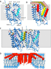
Models for the association of substrate, TatA, and TatB with TatC and for a TatA oligomer. Homology-based models for pea chloroplast TatC (blue), TatB TM (yellow), and TatA TM (red) based on structures of A. aeolicus TatC and E. coli TatB and TatA are shown. A, TatC is labeled for the cis (stroma) 1–4 exposed loops and tails, the TMs 1–6, and the trans (lumen) 1–3 loops. The pivotal C-proximal helix of TM5 is colored cyan. Residues of cis 1 and 2 important for RR binding are colored orange and red to designate loss of binding upon mutation to alanine, with red also designating essential glutamate residues that may coordinate the arginine guanidinium groups, and green designating residues that direct disulfide cross-linking to RR proximal residues of the signal peptide (56). B, arrangement of TatB (37, 45, 46) and TatA (54) with TatC TM5 under non-transporting conditions (see text). The patterned cylinders represent the APH segments and are approximately placed based on cross-linking. Docking of TatA on the Gln-234 (magenta) of TM4 in the translocase is proposed to initiate TatA oligomerization to form the pore (37, 54). C, a head-to-tail arrangement of two TatC subunits can explain how the signal peptide RR domain binds to TatC cis 1 and cis 2 regions and the H domain binds the TatB TM. In the panel, the signal peptide is hand-sketched from the RR through the helical H domain and is continuous with the early mature domain that is designated by the dashed line. D, a model of the TatA nonamer in detergent micelles adapted from Ref. (Protein Data Bank (PDB) accession 2LZS). The curved APH domains likely reflect the curvature of the micelle surface. The nonamer structure overlays a sketch of a lipid bilayer deformed by hydrophobic mismatch into a lipid half-pore based on ideas presented in Refs. and .
FIGURE 4.
FIGURE 4.
Proposed arrangement of TatABC units and bound substrate in the multimeric receptor complex. Graphic models of TatB (yellow), TatA (red), TatC (blue), and substrate (gray/black) are viewed from the cis compartment. The arrangements are based on cross-linking among Tat components and the substrate signal peptide and early mature domain. TatC TMs are labeled 1–6 on the top of the TMs, and only the TM helices of TatA and TatB are shown, except in C, which shows N-terminal tails of TatB, which are known to collectively cross-link. A and B, the face-to-face arrangement of dimers and tetramers is based on disulfide cross-linking, photocross-linking, and bismaleimide cross-linking (46, 54, 56) of TatC to TatC and photocross-linking between TatB and TatC (46). C and D, the arrangement of the substrate-bound TatABC is based on signal peptide cross-linking to TatC (20, 21, 47, 54, 56) and TatB, and the decrease of certain disulfide cross-links when signal peptide is bound. The signal peptide is depicted as a helical black line, the early mature domain is depicted as a dashed line, and the folded domain of the substrate is depicted as a sphere. Note that substrate binding increases the size of the chamber created by TatBC units where the TatA pore is proposed to assemble. It is also apparent that TatB controls TatA entry into the chamber. See the text for additional detail.

References

    1. Cline K., Theg S. (2007) The Sec and Tat protein translocation pathways in chloroplasts. in The Enzymes: Molecular Machines Involved in Protein Transport across Cellular Membranes (Dalbey R. E., Koehler C. M., Tamanoi F., eds), pp. 463–492, Academic Press, New York
    1. Palmer T., Berks B. C. (2012) The twin-arginine translocation (Tat) protein export pathway. Nat. Rev. Microbiol. 10, 483–496 - PubMed
    1. Fröbel J., Rose P., Müller M. (2012) Twin-arginine-dependent translocation of folded proteins. Philos. Trans. R. Soc. Lond. B Biol. Sci. 367, 1029–1046 - PMC - PubMed
    1. Celedon J. M., Cline K. (2013) Intra-plastid protein trafficking: how plant cells adapted prokaryotic mechanisms to the eukaryotic condition. Biochim. Biophys. Acta 1833, 341–351 - PMC - PubMed
    1. Berks B. C. (2015) The twin-arginine protein translocation pathway. Annu. Rev. Biochem., 84, 843–864 - PubMed

MeSH terms

Substances

Associated data

LinkOut - more resources