Observational Study
. 2015 Sep 15;121(18):3240-51.
doi: 10.1002/cncr.29455. Epub 2015 May 14.

Subclassification of prostate cancer circulating tumor cells by nuclear size reveals very small nuclear circulating tumor cells in patients with visceral metastases

Affiliations
Observational Study

Subclassification of prostate cancer circulating tumor cells by nuclear size reveals very small nuclear circulating tumor cells in patients with visceral metastases

Jie-Fu Chen et al. Cancer. .

Abstract

Background: Although enumeration of circulating tumor cells (CTCs) has shown some clinical value, the pool of CTCs contains a mixture of cells that contains additional information that can be extracted. The authors subclassified CTCs by shape features focusing on nuclear size and related this with clinical information.

Methods: A total of 148 blood samples were obtained from 57 patients with prostate cancer across the spectrum of metastatic states: no metastasis, nonvisceral metastasis, and visceral metastasis. CTCs captured and enumerated on NanoVelcro Chips (CytoLumina, Los Angeles, Calif) were subjected to pathologic review including nuclear size. The distribution of nuclear size was analyzed using a Gaussian mixture model. Correlations were made between CTC subpopulations and metastatic status.

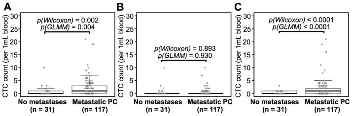
Results: Statistical modeling of nuclear size distribution revealed 3 distinct subpopulations: large nuclear CTCs, small nuclear CTCs, and very small nuclear CTCs (vsnCTCs). Small nuclear CTCs and vsnCTC identified those patients with metastatic disease. However, vsnCTC counts alone were found to be elevated in patients with visceral metastases when compared with those without (0.36 ± 0.69 vs 1.95 ± 3.77 cells/mL blood; P<.001). Serial enumeration studies suggested the emergence of vsnCTCs occurred before the detection of visceral metastases.

Conclusions: There are morphologic subsets of CTCs that can be identified by fundamental pathologic approaches, such as nuclear size measurement. The results of this observational study strongly suggest that CTCs contain relevant information regarding disease status. In particular, the detection of vsnCTCs was found to be correlated with the presence of visceral metastases and should be formally explored as a putative blood-borne biomarker to identify patients at risk of developing this clinical evolution of prostate cancer.

Keywords: NanoVelcro Chips; circulating tumor cells; nuclear size; prostate cancer; visceral metastasis.


Conflict of interest statement

Disclosure of Potential Conflicts of Interest: No potential conflicts of interest were disclosed.

Figures

Figure 1
Figure 1. The study design for identifying very-small-nuclear CTCs (vsnCTCs)
PC patients were recruited for CTC enumeration studies and each sample was placed into one of three cohorts depending on the metastatic status of the patient at the time of collection including: visceral metastasis, non-visceral (osseous/lymph node) metastasis, and non-metastatic disease. CTC enumeration studies were performed using highly sensitive NanoVelcro Chips in conjunction with the use of fluorescence microscopy. The identified CTCs (DAPI+/CK+/CD45-) were then subjected to nuclear size measurements. Through biostatistical analysis and modeling, we identified three subpopulations of CTCs: large-nuclear CTCs (lnCTCs), small-nuclear CTCs (snCTCs) and very-small-nuclear CTCs (vsnCTCs). The emergence of vsnCTCs correlates with the onset or presence of visceral metastases and may serve as the basis for a new diagnostic assay targeting the most aggressive form of PC. (Scale bars in the figures indicate 10 μm.)
Figure 2
Figure 2. Statistical analysis and modeling of CTC nuclear size distribution and CTC subpopulations
A) The histogram depicts the distribution of 304 CTCs according to their nuclear sizes. The black solid line shows the density of the optimal Gaussian mixture model (GMM) that best fits the histogram. The red, blue and grey solid lines represent the three cluster-specific Gaussian density curves. The dash lines indicate the cutoffs of our classification rule: assigning a sample to the cluster with the maximum Gaussian likelihood. B) The blue line shows the AIC values and the red line shows the BIC values for K (cluster number) from 1 to 4. The best cluster number is 3 as both AIC and BIC reached the minimum when K = 3.
Figure 3
Figure 3. Relationship between CTC nuclear sizes and metastatic status
A) Representative images of a lnCTC, a snCTC and a vsnCTC taken by fluorescence microscopy. The cells are stained with DAPI (blue), Alexa Fluor 488-conjugated anti-CK (green), and Alexa Fluor 555-conjugated anti-CD45 (orange). Histograms and cluster-specific Gaussian density curves were plotted using CTC counts versus nuclear sizes and classified into three metastatic statuses: B) No metastasis, in which lnCTCs account for the largest proportion (62%) among all the CTCs; C) Non-visceral metastasis, in which snCTCs constitute the major subpopulation (51%), followed by vsnCTCs (27%); and D) visceral metastasis, in which vsnCTCs account for the largest proportion of cells (65%), followed by snCTCs (20%). The proportions of the three CTC subpopulations varied significantly between different metastatic statuses as demonstrated by a two-sided χ2 test (p = 2.29×10⁻¹³).
Figure 4
Figure 4. Correlation between snCTC + vsnCTC counts and metastatic PC
Box plots are shown for A) total CTC counts, B) lnCTC counts, and C) snCTC + vsnCTC counts in 1.0 mL of blood. Boxes represent interquartile range, and the horizontal line across each box indicates median value. The y-axis represents CTC counts per 1.0 mL of blood. There are 31 enumerations from the category of no metastasis and 117 from metastatic PC patients (Supporting Figure 4). The data showed that the snCTC + vsnCTC counts obtained by using these nuclear size-based CTC definitions provide more information on metastatic status in PC than total CTC count. Statistical analyses were performed using two-sided Wilcoxon tests and Wald tests following a Generalized Linear Mixed Model (GLMM). All the p values are indicated in the figures.
Figure 5
Figure 5. Correlation between vsnCTC counts and aggressive PC with visceral metastasis
Box plots are shown for A) snCTC + vsnCTC counts, B) snCTC counts, and C) vsnCTC counts in 1.0 mL of blood. Boxes represent interquartile range, and the horizontal line across each box indicates median value. The y-axis represents CTC counts per 1.0 mL of blood. There are 44 enumerations from the category of non-visceral metastasis and 61 from the category of visceral metastasis (Supporting Figure 4). The results indicated that vsnCTCs rather than snCTCs are correlated with the presence of visceral metastases. Statistical analyses were performed using two-sided Wilcoxon tests and Wald tests following a Generalized Linear Mixed Model (GLMM). All the p values are indicated in the figures.
Figure 6
Figure 6. Summary of the 15 patients with visceral metastases
The beginning of each timeline (Day 0) indicates the time of first CTC enumeration by NanoVelcro Chips. The end of each timeline is the time of the end of follow-up. There are 3 patients (1117, 1161, 3701) initially presented with osseous/nodal metastases and later developed visceral lesions. vsnCTCs were found in these patients prior to radiographic detection of visceral metastases with the lead time ranging from 104 to 196 days (typical example see patient A in Supporting Figure 5A). Detailed clinical history of two other example cases (patient B and C) and observations on their vsnCTCs were presented in Supporting Figure 5B and 6.

Comment in

References

    1. Siegel R, Ma J, Zou Z, Jemal A. Cancer statistics, 2014. CA Cancer J Clin. 2014;64:9–29. - PubMed
    1. Pezaro CJ, Omlin A, Lorente D, et al. Visceral disease in castration-resistant prostate cancer. Eur Urol. 2014;65:270–273. - PMC - PubMed
    1. Gandaglia G, Karakiewicz PI, Briganti A, et al. Impact of the Site of Metastases on Survival in Patients with Metastatic Prostate Cancer. Eur Urol. 2014 - PubMed
    1. Pond GR, Sonpavde G, de Wit R, Eisenberger MA, Tannock IF, Armstrong AJ. The prognostic importance of metastatic site in men with metastatic castration-resistant prostate cancer. Eur Urol. 2014;65:3–6. - PubMed
    1. Halabi S, Kelly WK, Zhou H, et al. The site of visceral metastases (mets) to predict overall survival (OS) in castration-resistant prostate cancer (CRPC) patients (pts): A meta-analysis of five phase III trials. J Clin Oncol. 2014;5s:2014. (suppl; abstr 5002)

Publication types

MeSH terms