

Emerging optical methods for surveillance of Barrett's oesophagus
- PMID: 25975605
- PMCID: PMC5019028
- DOI: 10.1136/gutjnl-2013-306706
Emerging optical methods for surveillance of Barrett's oesophagus
Abstract
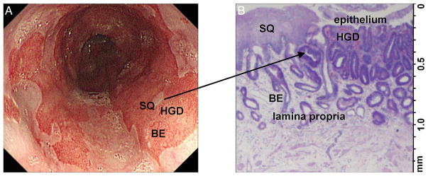
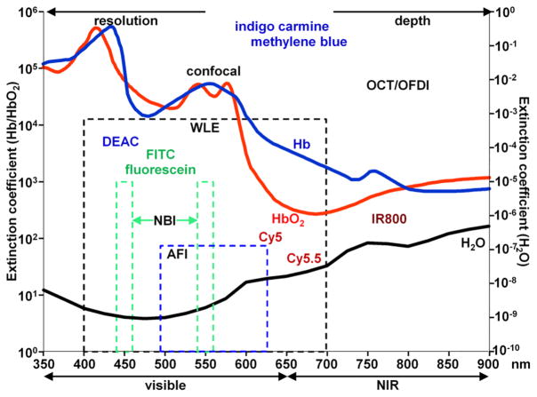
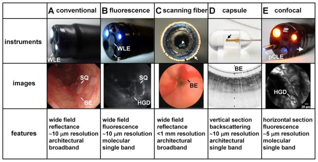
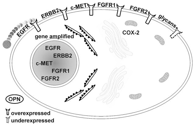
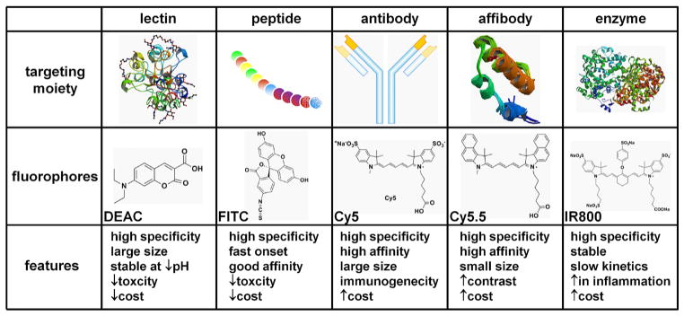
The rapid rise in incidence of oesophageal adenocarcinoma has motivated the need for improved methods for surveillance of Barrett's oesophagus. Early neoplasia is flat in morphology and patchy in distribution and is difficult to detect with conventional white light endoscopy (WLE). Light offers numerous advantages for rapidly visualising the oesophagus, and advanced optical methods are being developed for wide-field and cross-sectional imaging to guide tissue biopsy and stage early neoplasia, respectively. We review key features of these promising methods and address their potential to improve detection of Barrett's neoplasia. The clinical performance of key advanced imaging technologies is reviewed, including (1) wide-field methods, such as high-definition WLE, chromoendoscopy, narrow-band imaging, autofluorescence and trimodal imaging and (2) cross-sectional techniques, such as optical coherence tomography, optical frequency domain imaging and confocal laser endomicroscopy. Some of these instruments are being adapted for molecular imaging to detect specific biological targets that are overexpressed in Barrett's neoplasia. Gene expression profiles are being used to identify early targets that appear before morphological changes can be visualised with white light. These targets are detected in vivo using exogenous probes, such as lectins, peptides, antibodies, affibodies and activatable enzymes that are labelled with fluorescence dyes to produce high contrast images. This emerging approach has potential to provide a 'red flag' to identify regions of premalignant mucosa, outline disease margins and guide therapy based on the underlying molecular mechanisms of cancer progression.
Keywords: BARRETT'S OESOPHAGUS; IMAGING.
Published by the BMJ Publishing Group Limited. For permission to use (where not already granted under a licence) please go to http://group.bmj.com/group/rights-licensing/permissions.
Conflict of interest statement
Conflicts of Interest: None declared.
Figures
References
-
- Ferlay J, Soerjomataram I, Ervik M, et al. GLOBOCAN 2012 v1.0, Cancer Incidence and Mortality Worldwide: IARC CancerBase No. 11. Lyon, France: International Agency for Research on Cancer; 2013. http://globocan.iarc.fr.
-
- American Cancer Society. Cancer facts & figures 2012. Atlanta: American Cancer Society; 2012.
-
- Lagergren J, Bergström R, Lindgren A, et al. Symptomatic gastroesophageal reflux as a risk factor for esophageal adenocarcinoma. N Engl J Med. 1999;340:825–31. - PubMed
-
- Ryan AM, Duong M, Healy L, et al. Obesity, metabolic syndrome and esophageal adenocarcinoma: Epidemiology, etiology and new targets. Cancer Epidemiol. 2011;35:309–19. - PubMed
Publication types
MeSH terms
Supplementary concepts
Grants and funding
LinkOut - more resources
Full Text Sources
Other Literature Sources
Medical