. 2015 May 15;17(1):126.
doi: 10.1186/s13075-015-0647-9.

Evidence for cadherin-11 cleavage in the synovium and partial characterization of its mechanism

Affiliations

Evidence for cadherin-11 cleavage in the synovium and partial characterization of its mechanism

Erika H Noss et al. Arthritis Res Ther. .

Abstract

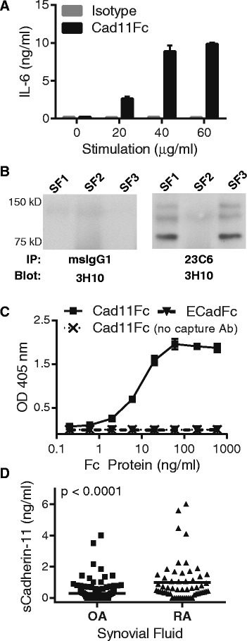
Introduction: Engagement of the homotypic cell-to-cell adhesion molecule cadherin-11 on rheumatoid arthritis (RA) synovial fibroblasts with a chimeric molecule containing the cadherin-11 extracellular binding domain stimulated cytokine, chemokine, and matrix metalloproteinases (MMP) release, implicating cadherin-11 signaling in RA pathogenesis. The objective of this study was to determine if cadherin-11 extracellular domain fragments are found inside the joint and if a physiologic synovial fibroblast cleavage pathway releases those fragments.

Methods: Cadherin-11 cleavage fragments were detected by western blot in cell media or lysates. Cleavage was interrupted using chemical inhibitors or short-interfering RNA (siRNA) gene silencing. The amount of cadherin-11 fragments in synovial fluid was measured by western blot and ELISA.

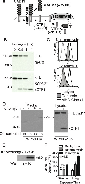
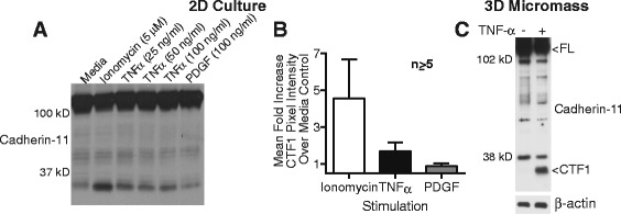
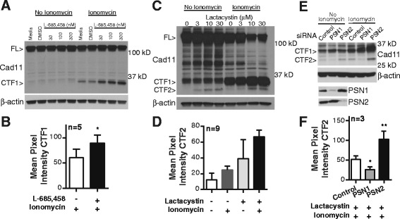
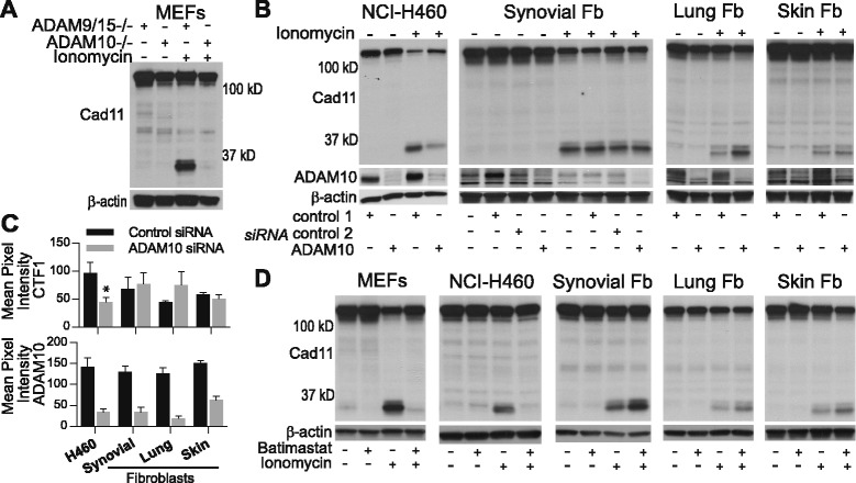
Results: Soluble cadherin-11 extracellular fragments were detected in human synovial fluid at significantly higher levels in RA samples compared to osteoarthritis (OA) samples. A cadherin-11 N-terminal extracellular binding domain fragment was shed from synovial fibroblasts after ionomycin stimulation, followed by presenilin 1 (PSN1)-dependent regulated intramembrane proteolysis of the retained membrane-bound C-terminal fragments. In addition to ionomycin-induced calcium flux, tumor necrosis factor (TNF)-α also stimulated cleavage in both two- and three-dimensional fibroblast cultures. Although cadherin-11 extracellular domains were shed by a disintegrin and metalloproteinase (ADAM) 10 in several cell types, a novel ADAM- and metalloproteinase-independent activity mediated shedding in primary human fibroblasts.

Conclusions: Cadherin-11 undergoes ectodomain shedding followed by regulated intramembrane proteolysis in synovial fibroblasts, triggered by a novel sheddase that generates extracelluar cadherin-11 fragments. Cadherin-11 fragments were enriched in RA synovial fluid, suggesting they may be a marker of synovial burden and may function to modify cadherin-11 interactions between synovial fibroblasts.


Figures

Fig. 1
Fig. 1
Evidence for cadherin-11 extracellular domain shedding in osteoarthritis (OA) and rheumatoid arthritis (RA) synovium. (a) Serum-starved RA synovial fibroblasts were incubated overnight with increasing concentrations of cad11Fc or isotype control antibody. IL-6 release was then measured by ELISA. Similar results were previously published and further characterized as discussed [3]. (b) Synovial fluid samples from OA patients were immunoprecipitated with a cadherin-11 extracellular domain antibody (23C6) followed by western blot analysis with a distinct cadherin-11 antibody (3H10). (c) Detection of the chimeric proteins cadherin-11-Fc or E-cadherin-Fc was measured using an ELISA developed to recognize the human cadherin-11 extracellular domain. The ELISA capture antibody was removed from the assay to serve as a negative control. The ELISA uses two anti-cadherin-11 monoclonal antibodies (23C6 and 3H10) recognizing distinct extracellular epitopes. (d) Soluble cadherin-11 fragments were detected by cadherin-11 extracellular domain specific ELISA in OA (n = 143, mean +/− standard deviation 0.28 +/− 0.56 ng/ml) and RA (n = 57, mean +/− standard deviation 0.99 +/− 1.3 ng/ml) synovial fluid patient samples. Cadherin-11 levels were significantly higher in RA synovial fluid (P <0.0001, two-tailed Student t-test)
Fig. 2
Fig. 2
Cadherin-11 cleavage stimulation by ionomycin. (a) Proposed cadherin-11 cleavage model shows that cadherin-11 is first cleaved extracellularly at the plasma membrane by a cell sheddase, generating a C-terminal fragment 1 (CTF1) and an extracellular domain fragment (sCad11). Then CTF1 is cleaved near the intracellular plasma membrane, releasing C-terminal fragment 2 (CTF2) into the cytosol. CTF2 is likely rapidly degraded by the proteasome but may also effect gene transcription. (b) Lysates from RA synovial fibroblasts stimulated with 5 μM ionomycin as indicated were analyzed by western blot using antibodies directed against cadherin-11 extracellular (3H10) or intracellular (5B2H5) epitopes (FL, full length cadherin-11). (c) Surface staining for cadherin-11 (3H10), MHC class I (W632) or isotype control on RA synovial fibroblasts before or after 1 hour ionomycin stimulation was determined by flow cytometry (representative, 2 experiments). (d) Culture media and cell lysates harvested from RA synovial fibroblasts treated with or without ionomycin for 1 hour were analyzed by western blot for extracellular (3H10) and intracellular (5B2H5) cadherin-11 epitopes. Culture media was left unconcentrated or concentrated approximately 12-fold (representative 3 experiments). (e) Culture media from unstimulated cells was immunoprecipitated with anti-cadherin-11 antibody 23C6 or appropriate isotype control prior to western blot analysis (representative 3 experiments). (f) CTF1 levels in twelve sequential experiments were assayed by measuring band pixel intensity inunstimulated and ionomycin-stimulated cell lysates by western blot using standard and longer exposure times. (Pooled, 6 cells lines. Statistical comparison to background by t-test. n.s. = not significantly different; * p<0.001).
Fig. 3
Fig. 3
Stimulation of cadherin-11 cleavage by TNF-α. (a) Lysates from rheumatoid arthritis (RA) synovial fibroblasts left untreated or treated with ionomycin, TNF-α, or platelet-derived growth factor (PDGF)-BB for 2 hours were assayed for cadherin-11 cleavage using monoclonal antibody 5B2H5. (b) Mean fold increase in C-terminal fragment 1 (CTF1) pixel intensity for the indicated stimulation over unstimulated controls was calculated across several experiments using RA fibroblasts from different donors (fold change over media control (mean +/− standard error of mean): ionomycin (n = 7, 4 cell lines) 4.6 +/− 2.1; TNF-α 25 ng/ml (n = 74 cell lines) 1.7 +/− 0.17; PDGF-BB 100 ng/ml (n = 53 cell lines) 0.89 +/− 0.074). (c) Synovial fibroblast micromasses were cultured for 21 days, transferred to media containing 0.2 % serum for 24 hours, and then stimulated for 5 days with or without TNF-α (10 ng/ml). Cadherin-11 cleavage was then assessed using a rabbit polyclonal antibody against the cadherin-11 cytoplasmic domain (representative of at least three separate experiments with two cell lines). Equal protein loading was confirmed by β-actin staining
Fig. 4
Fig. 4
Involvement of γ-secretase in cadherin-11 cleavage. (a) Cell lysates from synovial fibroblasts incubated overnight increasing concentrations of the γ-secretase inhibitor L-685-458 and then stimulated as indicated were analyzed for cadherin-11 cleavage (representative 5 experiments with 4 cells lines, full-length cadherin-11 (FL); cadherin-11 (Cad11)). (b) CTF1 nean pixel intensity was calculated in lysates from ionomycin-stimulated synovial fibroblasts pretreated with or without L-685-458 (dose range 300–3000 nM, * p=0.0304 by paired t-test, error bars reflect standard error of the mean). (c) Cell lysates from RA synovial fibroblasts incubated overnight with increasing concentrations of the proteosome inhibitor lactacystin and then stimulated as indicated were analyzed for cadherin-11 cleavage (representative of 9 experiments with 5 cells lines). (d) CTF2 mean pixel intensity was calculated in lysates from synovial fibroblast treated with or without ionomycin (5 μM) and lactacystin (3–10 μM) as indicated (p<0.0001 by one-way ANOVA, error bars reflect standard error of the mean). (e) RA synovial fibroblasts were transfected with the indicated siRNAs, incubated overnight with 10 μM lactacystin, and then were analyzed for cadherin-11 cleavage in the presence or absence of ionomycin (representative 3 experiments with 3 cell lines). Silencing was confirmed by western blot. β-actin levels were used fro protein loading control. (f) Mean pixel intensity was calculated for CTF2 bands in siRNA-transfected RA synovial fibroblasts pretreated with 10 μM lactacystin and then stimulated with ionomycin (* p=0.0045, paired t-test; **p=0.0136 paired t-test, n=3, error bars reflect standard deviation of the mean).
Fig. 5
Fig. 5
Comparison of sheddase activity in mouse embryonic fibroblasts (MEFs), NCI-H460, and primary human fibroblasts. (a) Lysates from MEFs genetically deficient in a disintegrin and metalloproteinase (ADAM10 −/−) or (b) from NCI-H460, synovial fibroblasts, lung fibroblasts, or skin fibroblasts transfected with control or ADAM10 siRNA were analyzed for cadherin-11 cleavage in the presence or absence of ionomycin (representative of at least three experiments, fibroblasts (Fb). ADAM10 siRNA silencing was confirmed by western blot and β-actin levels confirmed equal protein loading. (c) C-terminal fragment 1 (CTF1) and ADAM10 expression was measured by calculating the mean pixel intensity of CTF1 and ADAM10 bands across several experiments in control and ADAM10 siRNA treated cells for the indicated cell types (H460, n = 3; * P = 0.049 by paired t-test; lung and skin fibroblasts, n = 3; synovial fibroblasts, n = 6, five separate rheumatoid arthritis (RA) lines; error bars reflect standard error of mean). ADAM10 silencing efficiency (mean+/−standard deviation): H460 cells, 77.0 + 5.67%; synovial fibroblasts, 70.2+/−25.5%; lung fibroblast, 85.0+/−8.04%; skin fibroblasts, 58.8+/−7.77%. (d) Cell lysates from indicated cells treated overnight with the metalloproteinase inhibitor batimastat (10 μM) or appropriate dimethyl sulfoxide (DMSO) vehicle control were assayed for cadherin-11 cleavage in the presence and absence of ionomycin stimulation (representative of at least three experiments per cell type). Equal protein loading was confirmed by β-actin staining

References

    1. Bottini N, Firestein GS. Duality of fibroblast-like synoviocytes in RA: passive responders and imprinted aggressors. Nat Rev Rheumatol. 2013;9:24–33. doi: 10.1038/nrrheum.2012.190. - DOI - PMC - PubMed
    1. Noss EH, Brenner MB. The role and therapeutic implications of fibroblast-like synoviocytes in inflammation and cartilage erosion in rheumatoid arthritis. Immunol Rev. 2008;223:252–70. doi: 10.1111/j.1600-065X.2008.00648.x. - DOI - PubMed
    1. Chang SK, Noss EH, Chen M, Gu Z, Townsend K, Grenha R, et al. Cadherin-11 regulates fibroblast inflammation. Proc Natl Acad Sci U S A. 2011;108:8402–7. doi: 10.1073/pnas.1019437108. - DOI - PMC - PubMed
    1. Kiener HP, Lee DM, Agarwal SK, Brenner MB. Cadherin-11 induces rheumatoid arthritis fibroblast-like synoviocytes to form lining layers in vitro. Am J Pathol. 2006;168:1486–99. doi: 10.2353/ajpath.2006.050999. - DOI - PMC - PubMed
    1. Kiener HP, Niederreiter B, Lee DM, Jimenez-Boj E, Smolen JS, Brenner MB. Cadherin 11 promotes invasive behavior of fibroblast-like synoviocytes. Arthritis Rheum. 2009;60:1305–10. doi: 10.1002/art.24453. - DOI - PMC - PubMed

Publication types