

doi: 10.3324/haematol.2015.126870.
Epub 2015 May 14.
Mice are poor heme absorbers and do not require intestinal Hmox1 for dietary heme iron assimilation
Affiliations
- PMID: 25975840
- PMCID: PMC4800685
- DOI: 10.3324/haematol.2015.126870
Item in Clipboard
Mice are poor heme absorbers and do not require intestinal Hmox1 for dietary heme iron assimilation
Haematologica.
2015 Sep.
No abstract available
Keywords: dietary heme iron assimilation; high-heme diet; intestinal Hmox1; iron overload; mice.
Figures
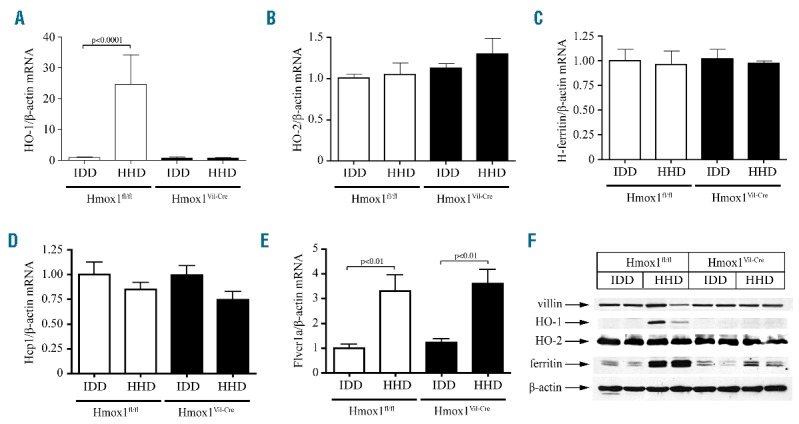
Effects of dietary heme excess in expression of intestinal heme and iron metabolism genes. 6-week old male Hmox1fl/fl and Hmox1Vil-Cre mice (n=5 for each genotype) were fed for two weeks with an IDD (containing only 2–6 ppm iron) or a HHD (IDD supplemented with 30 mmol/kg hemin, equivalent to 2% carbonyl iron in terms of iron quantity). At the end point the animals were sacrificed and duodenal RNA and protein extracts were prepared for qPCR and Western blot analysis, respectively. (A) The HHD promoted 23-fold induction of intestinal HO-1 mRNA in control Hmox1fl/fl but not in Hmox1Vil-Cre mice. (B–D) Intestinal HO-2 (B), H-ferritin (C) and Hcp1 (D) mRNA levels were not significantly affected by the diets and the genotypes. Constitutive expression of HO-2 mRNA was similar to that of HHD-induced HO-1 mRNA in Hmox1fl/fl mice. (E) The HHD stimulated intestinal Flvcr1a mRNA expression in both Hmox1fl/fl and Hmox1Vil-Cre mice. qPCR data in (A–E) are presented as the mean ± SEM. Statistical analysis was performed by one-way ANOVA. (F) The HHD induced intestinal HO-1 expression in control Hmox1fl/fl mice and intestinal ferritin expression in both Hmox1fl/fl and Hmox1Vil-Cre mice. Expression of HO-2, villin (intestinal marker) and β-actin (loading control) was not affected by the diets and the genotypes. Data from 2 representative mice are shown. Similar results were obtained with samples from the other mice used in the experiment.
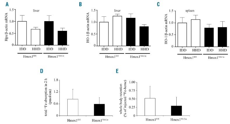
Effects of dietary heme excess in expression of hepatic and splenic heme metabolism genes, and analysis of intestinal heme transport. (A–C) Hepatic and splenic RNA was prepared from the mice described in Figure 1 and used for qPCR analysis. Dietary heme excess did not induce hemopexin (Hpx) mRNA in the liver (A), and HO-2 mRNA in the liver (B) or spleen (C). qPCR data are presented as the mean ± SEM. Statistical analysis was performed by one-way ANOVA. (D and E) Disruption of intestinal HO-1 did not affect absorption of 59Fe from 59Fe-Hb. A murine hemolysate containing 59Fe-Hb was administered to ligated intestinal loops from Hmox1fl/fl (n=5) and Hmox1Vil-Cre mice (n=6) for 2 h. (D) Total intestinal absorption of 59Fe. (E) Retention of 59Fe in the whole carcass (excluding the intestine). Data are presented as the mean ± SEM. Statistical analysis was performed by two-tailed Student’s t test.
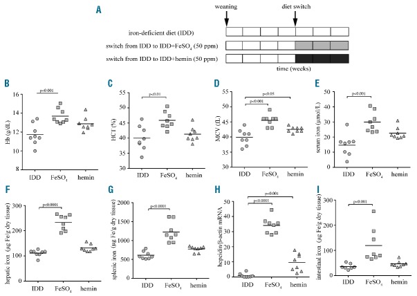
Iron deficiency anemia is efficiently corrected by a diet supplemented with inorganic iron but not heme, due to low luminal heme transport in the gut. 3-week old female C57BL/6 mice (n=24) were fed with an IDD for four weeks. One group of the animals (n=8) remained on this diet for another three weeks; a second group (n=8) was switched to IDD supplemented with 50 ppm FeSO4 and a third group (n=8) was switched to IDD supplemented with 50 ppm hemin. At the end point, blood was prepared and used for hematologic and serum iron analysis, hepatic RNA was prepared for qPCR analysis, and tissues were used for assessment of total (non-heme and heme) iron content. (A) Schematic representation of experimental design. (B) Hb concentration. (C) Hematocrit (HCT). (D) Mean corpuscular volume (MCV). (E) Serum iron. (F) Hepatic iron. (G) Splenic iron. (H) Hepcidin mRNA was induced 34-fold by dietary FeSO4 and 9.5-fold by dietary hemin supplementation. (I) Intestinal iron levels were increased 3.3-fold following dietary FeSO4 supplementation and were not significantly affected by dietary hemin. All data are presented as the mean ± SEM. Statistical analysis was performed by one-way ANOVA.
Comment in
-
Dietary and pharmacological factors affecting iron absorption in mice and man (Comment for a Letter to the editor).Haematologica. 2016 Mar;101(3):e120-1. doi: 10.3324/haematol.2015.138255. Haematologica. 2016. PMID: 26928250 Free PMC article. No abstract available.
-
Response to: Dietary and pharmacological factors affecting iron absorption in mice and man.Haematologica. 2016 Mar;101(3):e122. doi: 10.3324/haematol.2015.139741. Haematologica. 2016. PMID: 26928251 Free PMC article. No abstract available.
References
Publication types
MeSH terms
Substances
Grants and funding
LinkOut - more resources
Full Text Sources
Other Literature Sources
Medical
