. 2016 Feb;17(2):261-71.
doi: 10.1111/mpp.12278. Epub 2015 Jun 18.

Antifungal activity of the ribosome-inactivating protein BE27 from sugar beet (Beta vulgaris L.) against the green mould Penicillium digitatum

Affiliations

Antifungal activity of the ribosome-inactivating protein BE27 from sugar beet (Beta vulgaris L.) against the green mould Penicillium digitatum

Lucía Citores et al. Mol Plant Pathol. 2016 Feb.

Abstract

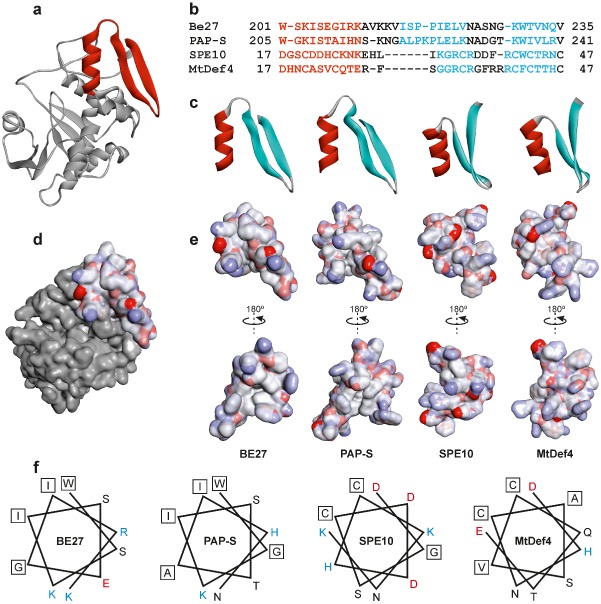
The ribosome-inactivating protein BE27 from sugar beet (Beta vulgaris L.) leaves is an apoplastic protein induced by signalling compounds, such as hydrogen peroxide and salicylic acid, which has been reported to be involved in defence against viruses. Here, we report that, at a concentration much lower than that present in the apoplast, BE27 displays antifungal activity against the green mould Penicillium digitatum, a necrotrophic fungus that colonizes wounds and grows in the inter- and intracellular spaces of the tissues of several edible plants. BE27 is able to enter into the cytosol and kill fungal cells, thus arresting the growth of the fungus. The mechanism of action seems to involve ribosomal RNA (rRNA) N-glycosylase activity on the sarcin-ricin loop of the major rRNA which inactivates irreversibly the fungal ribosomes, thus inhibiting protein synthesis. We compared the C-terminus of the BE27 structure with antifungal plant defensins and hypothesize that a structural motif composed of an α-helix and a β-hairpin, similar to the γ-core motif of defensins, might contribute to the specific interaction with the fungal plasma membranes, allowing the protein to enter into the cell.

Keywords: apoplast; defensin; green mould; plant defence; pokeweed antiviral protein (PAP); polynucleotide:adenosine glycosylase; rRNA N-glycosylase.


Figures

Figure 1
Figure 1
Antifungal activity of BE27 against Penicillium digitatum. Antifungal activity of BE27 against P. digitatum was measured in a microtitre plate bioassay. Conidia of P. digitatum were grown in potato dextrose broth (PDB) medium in the presence of different concentrations of BE27. Fungal growth was measured as the increase in absorbance at 620 nm. The curves represent buffer control (filled circles), 0.6 μg/mL BE27 (open circles), 4 μg/mL BE27 (filled squares) and 8 μg/mL BE27 (open squares). Conidia of P. digitatum were grown in PDB for 28 h before exposure to 8 μg/mL BE27 (filled triangles).
Figure 2
Figure 2
(a) Morphological changes in Penicillium digitatum mycelium exposed to BE27. Penicillium digitatum mycelium was grown in the absence (control) or presence of 5 μg/mL of BE27. After 24 h of incubation, samples were stained with calcofluor white (CFW) and visualized under the microscope. Panels represent bright‐field images (left) and fluorescence images indicative of CFW staining (right) for the same fields. Bar, 20 μm. (b) Interaction between BE27 and P. digitatum. To visualize the interaction of BE27 with fungal structures, conidia of P. digitatum were grown in vitro in potato dextrose broth (PDB) medium for 24 h before exposure to 5 μg/mL of CY3‐BE27 for 24 h. After washing, the samples were visualized with a confocal laser microscope. The red channel image (right panel) and the corresponding interference contrast image (left panel) are shown. Bar, 5 μm.
Figure 3
Figure 3
rRNAN‐glycosylase activity of BE27 on yeast and Penicillium digitatum ribosomes. Left: rRNAN‐glycosylase activity was assayed as indicated in Experimental procedures. Each lane contained 3 μg of RNA isolated from either untreated (control) or BE27‐treated ribosomes from yeast or P. digitatum. The arrows indicate the RNA fragments released as a consequence of ribosome‐inactivating protein (RIP) action on acid aniline treatment (+). Numbers indicate the size of the standards (M) in nucleotides. Right: sarcin–ricin loop of the large rRNA from yeast and Penicillium. The sequences from Saccharomyces cerevisiae (accession number J01355) and Penicillium solitum (JN642222) were downloaded from the National Center for Biotechnology Information (NCBI) sequence database (http://www.ncbi.nlm.nih.gov/nucleotide/). The large rRNA 3′ end from Penicillium was determined by alignment with the large rRNA from Saccharomyces. The adenine released by RIP action (bold type), the site of splitting by the acid aniline (arrow) and the size of the generated fragment are also indicated.
Figure 4
Figure 4
Antifungal and rRNAN‐glycosylase activity of BE27 against Penicillium digitatum. (a) Penicillium digitatum was grown in potato dextrose broth (PDB) in the absence (control) or presence of 4 μg/mL BE27 for 5 days. The mycelium was extensively washed with sterile water and harvested to extract the RNA and DNA. Representative photographs of two plates are shown. (b) rRNAN‐glycosylase activity was assayed as indicated in Experimental procedures. Each lane contained 3 μg of RNA isolated from either untreated (control) or BE27‐treated cultures from P. digitatum. For comparative purposes, yeast RNA depurinated by BE27 is also included in the assay. The arrows indicate the RNA fragments released as a consequence of ribosome‐inactivating protein (RIP) action on acid aniline treatment (+). Numbers indicate the size of the standards (M) in nucleotides. (c) The DNA was isolated as indicated in Experimental procedures and 3 μg was electrophoresed. The numbers indicate the corresponding size of the standards (λDNA HindIII/EcoRI) in Kb.
Figure 5
Figure 5
Structure of the α‐helix/γ‐core motif of BE27 compared with PAPS (pokeweed antiviral protein) and the defensins SPE10 and MtDef4. (a) Structure of BE27 indicating the position of the α‐helix/γ‐core motif from amino acid 201 to amino acid 235 (red ribbon). (b) Sequence alignments based on the three‐dimensional alignments using the program Expresso (Di Tommaso et al., 2011). Colouring of the alignments indicates the presence of α‐helix (red) or β‐sheet (cyan) secondary structures. (c) Ribbon representation of the α‐helix/γ‐core motif from BE27 expanding from amino acid 201 to amino acid 235 and their homologues from PAPS, SPE10 and MtDef4. The α‐helix and β‐strands are coloured in red and cyan, respectively. (d, e) Electrostatic surface of the α‐helix/γ‐core motif from BE27 localized in the whole protein (d) or compared with the same motif in PAPS, SPE10 and MtDef4 (e). Electrostatic potential is indicated in red (negative charge), white (neutral) and blue (positive charge). (f) Helical wheel drawing of the helices of BE27, PAPS, SPE10 and MtDef4. The amino acid charge is indicated in red (negative), black (neutral) and blue (positive). Hydrophobic amino acids are boxed.

Similar articles

Cited by

References

    1. Barbieri, L. , Valbonesi, P. , Bonora, E. , Gorini, P. , Bolognesi, A. and Stirpe, F. (1997) Polynucleotide:adenosine glycosidase activity of ribosome‐inactivating proteins: effect on DNA, RNA and poly(A). Nucleic Acids Res. 25, 518–522. - PMC - PubMed
    1. Barbieri, L. , Polito, L. , Bolognesi, A. , Ciani, M. , Pelosi, E. , Farini, V. , Jha, A.K. , Sharma, N. , Vivanco, J.M. , Chambery, A. , Parente, A. and Stirpe, F. (2006) Ribosome‐inactivating proteins in edible plants and purification and characterization of a new ribosome‐inactivating protein from Cucurbita moschata . Biochim. Biophys. Acta, 1760, 783–792. - PubMed
    1. Baykal, U. and Tumer, N.E. (2007) The C‐terminus of pokeweed antiviral protein has distinct roles in transport to the cytosol, ribosome depurination and cytotoxicity. Plant J. 49, 995–1007. - PubMed
    1. Becker, N. and Benhar, I. (2012) Antibody‐based immunotoxins for the treatment of cancer. Antibodies, 1, 39–69.
    1. Berglund, L. , Brunstedt, J. , Nielsen, K.K. , Chen, Z. , Mikkelsen, J.D. and Marcker, K.A. (1995) A proline‐rich chitinase from Beta vulgaris . Plant Mol. Biol. 27, 211–216. - PubMed

Publication types

MeSH terms

Associated data