. 2015 May 15;46(1):53.
doi: 10.1186/s13567-015-0194-z.

Invasion and persistence of Mycoplasma bovis in embryonic calf turbinate cells

Affiliations

Invasion and persistence of Mycoplasma bovis in embryonic calf turbinate cells

Sibylle Bürki et al. Vet Res. .

Abstract

Mycoplasma bovis is a wall-less bacterium causing bovine mycoplasmosis, a disease showing a broad range of clinical manifestations in cattle. It leads to enormous economic losses to the beef and dairy industries. Antibiotic treatments are not efficacious and currently no efficient vaccine is available. Moreover, mechanisms of pathogenicity of this bacterium are not clear, as few virulence attributes are known. Microscopic observations of necropsy material suggest the possibility of an intracellular stage of M. bovis. We used a combination of a gentamicin protection assay, a variety of chemical treatments to block mycoplasmas entry in eukaryotic cells, and fluorescence and transmission electron microscopy to investigate the intracellular life of M. bovis in calf turbinate cells. Our findings indicate that M. bovis invades and persists in primary embryonic calf turbinate cells. Moreover, M. bovis can multiply within these cells. The intracellular phase of M. bovis may represent a protective niche for this pathogen and contribute to its escape from the host's immune defense as well as avoidance of antimicrobial agents.


Figures
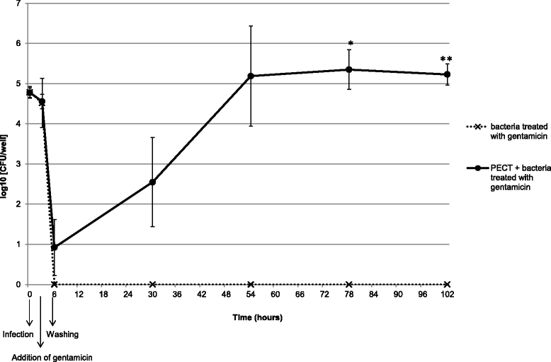
Figure 1
Figure 1
Intracellular growth curve of M. bovis in PECT cells. Broken line and crosses: M. bovis strain JF4278 with gentamicin. Continuous line and dots: PECT cells and M. bovis strain JF4278 with gentamicin. The data shown are the mean values of triplicates of 2 independent experiments and standard deviations of individual measurements are indicated as bars. *P < 0.05, **P < 0.01.
Figure 2
Figure 2
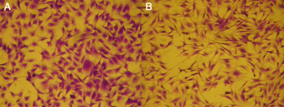
PECT cells morphology by light microscopy. Morphological features of PECT cells stained with crystal violet. Panel A: PECT cells at passage 8. Panel B: PECT cells at passage 10. Magnification 112.5 ×.
Figure 3
Figure 3
Infection model of M. bovis with PECT cells. Time points 0, 6 and 54 h post infection with an MOI between 2 and 30 are shown. Horizontally striped columns correspond to M. bovis alone in growth medium, while filled columns correspond to the infection of PECT cells. The data shown are the mean values of triplicates of 2 independent experiments. Standard deviations of individual measurements are indicated as bars. *P < 0.05, **P < 0.01, ***P < 0.001.
Figure 4
Figure 4
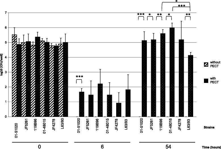
Gentamicin protection assay. Time points 0, 6 and 54 h post infection with an MOI between 2 and 30 are shown. Time 6 h post infection corresponds to the end of the gentamicin treatment. Diagonally striped columns correspond to M. bovis alone in growth medium supplemented with gentamicin, while dotted columns correspond to the infection of PECT cells with growth medium supplemented with gentamicin. The data shown are the mean values of triplicates of 2 independent experiments. Standard deviations of individual measurements are indicated as bars. *P < 0.05, **P < 0.01, ***P < 0.001.
Figure 5
Figure 5
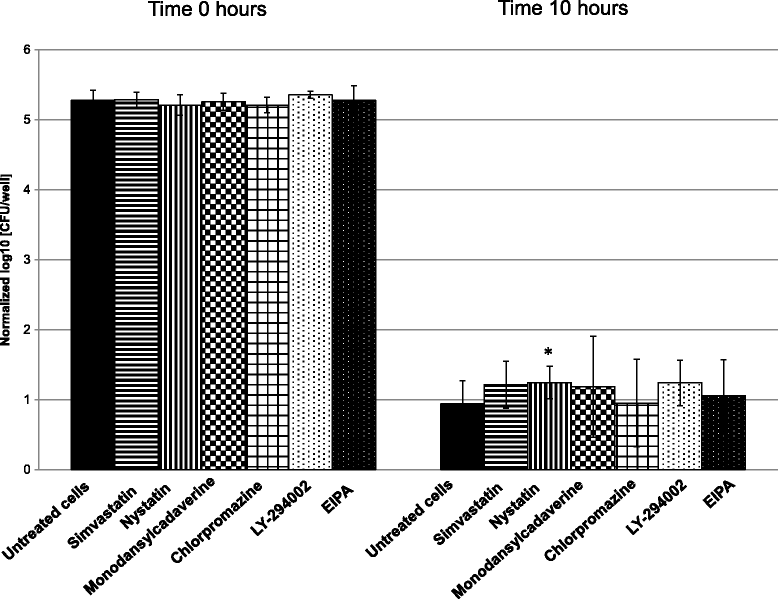
Inhibition of M. bovis entry in PECT cells by chemical treatment. The data shown are the mean values of the triplicates of 2 independent experiments at time 0 and 10 h using an MOI between 2 and 30, the latter 4 h after the gentamicin incubation time. Standard deviations of the individual measurements are indicated as bars. *P < 0.05.
Figure 6
Figure 6
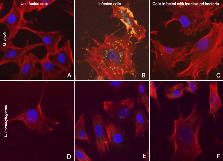
Differential fluorescence microscopy of PECT cells infection with M. bovis and L. monocytogenes. Panels A, B and C: infection with M. bovis at an MOI of 3400, 6 h post infection. Panels D, E and F: infection with L. monocytogenes at an MOI of 55, 3 h post infection. Panels A and D: negative controls. Panels B and E: infected cells. Panels C and F: cells infected with formaldehyde-inactivated bacteria. Nuclei are stained in blue, F-actin is stained in red, intracellular bacteria are stained in green and extracellular bacteria are stained in yellow-orange. Merged images. Magnification 600 ×.
Figure 7
Figure 7
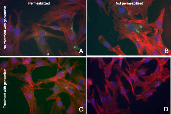
Fluorescence microscopy of the gentamicin protection assay with permeabilized and not permeabilized cells using antibodies directed against M. bovis . MOI of 10; 54 hours post infection. Panel A: Permeabilized PECT cells without gentamicin treatment infected with M. bovis JF4278. Mycoplasmas are visible intra- and extracellularly. Panel B: Non permeabilized PECT cells without gentamicin treatment infected with M. bovis JF4278. Only extracellular mycoplasmas are visible. Panel C: Permeabilized PECT cells with gentamicin treatment infected with M. bovis JF4278. Only intracellular mycoplasmas are visible. Panel D: Not permeabilized PECT cells with gentamicin treatment infected with M. bovis JF4278. No mycoplasmas are visible. Nuclei are stained in blue, F-actin is stained in red, mycoplasmas are stained in green. Merged images. Magnification 600 ×.
Figure 8
Figure 8
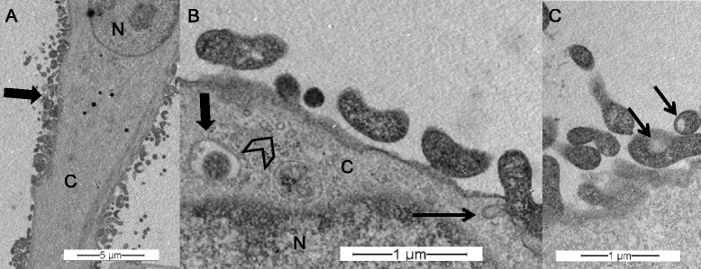
Transmission electron microscopy of PECT cells infected with M. bovis . MOI of 600, 16 h post infection. Panel A: M. bovis infected PECT cell, M. bovis clusters (bold arrow). Panel B: PECT cell invagination (thin arrow), intracellular vesicles (arrowhead), intracellular, membrane-bound M. bovis (bold arrow). Panel C: electron-lucent space in mycoplasmal cytoplasm (thin arrow). N: PECT cell nucleus; C: PECT cell cytoplasm.

Similar articles

Cited by

References

    1. Stipkovits L, Rosengarten R, Frey J. Mycoplasmas of ruminants: pathogenicity, diagnostics, epidemiology and molecular genetics. Brussels: European Commission; 1999.
    1. Caswell JL, Archambault M. Mycoplasma bovis pneumonia in cattle. Anim Health Res Rev. 2007;8:161–186. doi: 10.1017/S1466252307001351. - DOI - PubMed
    1. Gonzalez RN, Wilson DJ. Mycoplasmal mastitis in dairy herds. Vet Clin North Am Food Anim Pract. 2003;19:199–221. doi: 10.1016/S0749-0720(02)00076-2. - DOI - PubMed
    1. Pfützner H, Sachse K. Mycoplasma bovis as an agent of mastitis, pneumonia, arthritis and genital disorders in cattle. Rev Sci Tech. 1996;15:1477–1494. - PubMed
    1. Walz PH, Mullaney TP, Render JA, Walker RD, Mosser T, Baker JC. Otitis media in preweaned Holstein dairy calves in Michigan due to Mycoplasma bovis. J Vet Diagn Invest. 1997;9:250–254. doi: 10.1177/104063879700900305. - DOI - PubMed

Publication types

MeSH terms