. 2015 May 15;16(1):98.
doi: 10.1186/s13059-015-0666-5.

Visualizing translocation dynamics and nascent transcript errors in paused RNA polymerases in vivo

Affiliations

Visualizing translocation dynamics and nascent transcript errors in paused RNA polymerases in vivo

Masahiko Imashimizu et al. Genome Biol. .

Erratum in

Abstract

Background: Transcription elongation is frequently interrupted by pausing signals in DNA, with downstream effects on gene expression. Transcription errors also induce prolonged pausing, which can lead to a destabilized genome by interfering with DNA replication. Mechanisms of pausing associated with translocation blocks and misincorporation have been characterized in vitro, but not in vivo.

Results: We investigate the pausing pattern of RNA polymerase (RNAP) in Escherichia coli by a novel approach, combining native elongating transcript sequencing (NET-seq) with RNase footprinting of the transcripts (RNET-seq). We reveal that the G-dC base pair at the 5' end of the RNA-DNA hybrid interferes with RNAP translocation. The distance between the 5' G-dC base pair and the 3' end of RNA fluctuates over a three-nucleotide width. Thus, the G-dC base pair can induce pausing in post-translocated, pre-translocated, and backtracked states of RNAP. Additionally, a CpG sequence of the template DNA strand spanning the active site of RNAP inhibits elongation and induces G-to-A errors, which leads to backtracking of RNAP. Gre factors efficiently proofread the errors and rescue the backtracked complexes. We also find that pausing events are enriched in the 5' untranslated region and antisense transcription of mRNA genes and are reduced in rRNA genes.

Conclusions: In E. coli, robust transcriptional pausing involves RNAP interaction with G-dC at the upstream end of the RNA-DNA hybrid, which interferes with translocation. CpG DNA sequences induce transcriptional pausing and G-to-A errors.


Figures
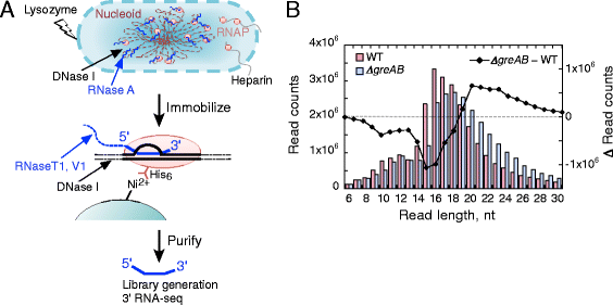
Fig. 1
Fig. 1
RNET-seq, the read-length-specific NET-seq approach for the analysis of transcriptional pausing and errors in vivo. (a) An overview of the preparation of the RNA samples for RNET-seq. The 3′ RNA transcripts protected by RNAP from RNases were isolated from E. coli cells. (b) Different distribution of read lengths between the WT and ΔgreAB cells
Fig. 2
Fig. 2
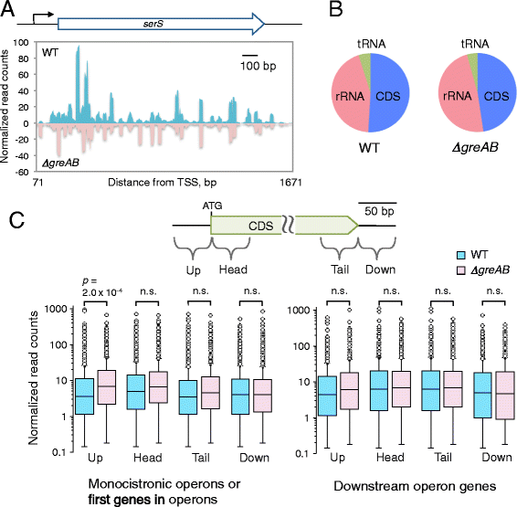
Comparison of genome-wide transcription in E. coli WT and ΔgreAB cells. (a) A transcription pausing profile of the serR gene. TSS transcription start site. (b) Mapped sequencing reads from paused RNAP complexes carrying mRNA (coding DNA sequence (CDS)), tRNA and rRNA. (c) GreAB proteins reduce pausing in 5′ UTRs of E. coli mRNA genes. Each box plot represents the quartile of normalized read counts in a 50-bp window for each gene body: upstream (Up), head, tail, and downstream (Down). mRNA genes with normalized read counts >0.1 (n = 1847 for left panel and n = 882 for right panel) were used for the analysis. The p-value of two-tailed t-test is shown for a pair with statistically significant difference between the WT and ΔgreAB data. The p-values >0.05 are labeled as non-significant (n.s.)
Fig. 3
Fig. 3
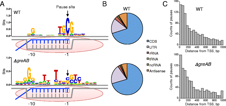
Transcription pausing detected by RNET-seq in E. coli WT and ΔgreAB cells. (a) Pause-inducing elements (PIEs) of the non-template DNA strand. Information content is represented by sequence logos [51]. Positions −1 and −10 of DNA (gray) correspond to the RNA (blue) 3′ and 5′ ends of the RNA-DNA hybrid within RNAP (pink oval) in pre-translocated ECs. The active site is shown as an empty square. P(0.9, 100) and P(0.9, 160) were used for WT (n = 758) and ΔgreAB (n =419), respectively (see main text for the parameters). A frequency matrix and MAP scores for the PIEs are shown in Table S1 in Additional file 2. (b) Categorization of all RNAP pauses by RNA type. The non-coding RNA (ncRNA) and antisense RNA were defined using the gene annotation file of E. coli (see “Materials and methods”). (c) Pausing frequently occurs in regions proximal to transcript start sites (TSS). For panels B and C, P(0.7, 100) is used in order to increase the number of samples. Note that even when using this reduced stringency the consensus sequence for pausing remains unaffected (Fig. S6B in Additional file 1)
Fig. 4
Fig. 4
RNAP pauses during antisense transcription detected by RNET-seq. (a) Scatter plot of antisense and sense transcription in the WT strain. Each dot represents a 50-bp “tail” (Fig. 2C) region of a gene. The Pearson’s correlation coefficient (r) is shown, and y = x (dotted line) represents a positive correlation. (b) Convergent transcription and pauses in the rfaH and tatD genes of WT cells. The reads mapped to plus and minus strands of genomic DNA are shown in blue and orange, respectively. The sequences for pause sites (labeled by 1–8) are shown
Fig. 5
Fig. 5
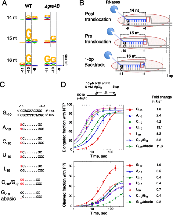
The G-dC base pair at the 5′ end of the RNA-DNA hybrid interferes with RNAP translocation in vivo and in vitro. (a) PIEs generated by the single-length RNET-seq analysis for 14-, 15- or 16-nt reads from WT and ΔgreAB cells. DNA positions −9, −10 and −11, where −1 corresponds to the 3′ RNA base, are shown. Pausing was defined by P(0.9, 50). The full-length PIEs are shown in Figs. S9 and S10 in Additional file 1. For 14-nt reads, the pause sties of mapqmean >10 are used (n = 286 and 258 for WT and ΔgreAB, respectively; Fig. S11 in Additional file 1). (b) Model for robust transcription pausing in the post- (14 nt), pre-translocated (15 nt) or 1-bp backtracked (16 nt) state according to the −9, −10 or −11 position of the riboG-dC. (c) Ten-nucleotide RNA strands (top) and the template DNA strands (TDS) in the ECs used for the biochemical assay. The RNA and template DNA bases, carrying sequence different from the original G−10 scaffold, are indicated in red. (d) Effects of different −10 and +1 bases on the elongation (upper) and pyrophosphorolysis (lower) of an EC carrying a 10-nt transcript (EC10). Reaction scheme is shown at the top. The apparent rate constants (k) for these two reactions were obtained by fitting the data to single-exponential curves. The mean values of two or three independent experiments ± standard deviations are shown. PPi pyrophosphate
Fig. 6
Fig. 6
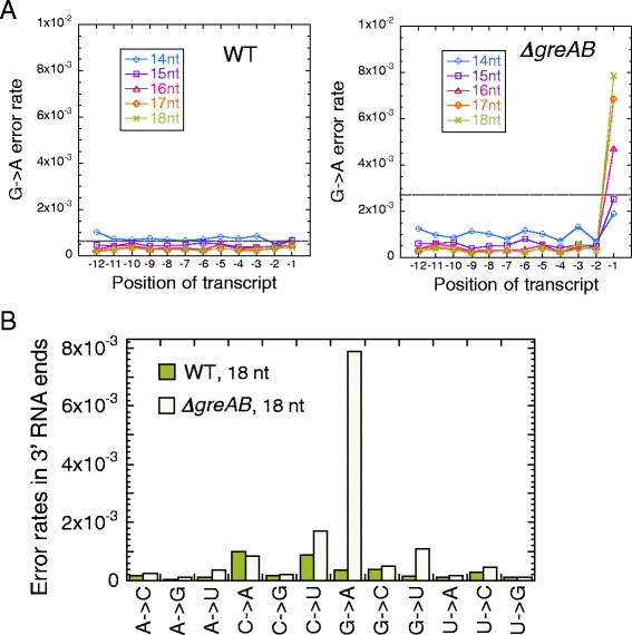
Transcriptional errors detected by single-length (14–18 nt) RNET-seq. (a) G-to-A error rates at the 3′ RNA ends are increased in the absence of Gre factors. Position −1 corresponds to the 3′ RNA end. Broken lines represent values for mean error rate + standard deviation in the −12 to −1 positions of the 14- to 18-nt reads. (b) Error rates in the 3′ ends of nascent transcripts detected by single-length (18 nt) RNET-seq
Fig. 7
Fig. 7
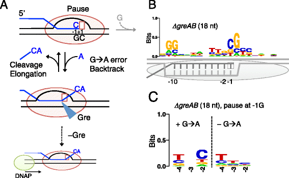
Transcriptional pauses and errors frequently occur at CpG sequences in vivo. (a) G-to-A error at the 3′ RNA end induces backtracking of RNAP in the C−1G+1 motif. These pauses are rescued and the errors are corrected by Gre factors. In the absence of Gre factors, the backtracked RNAP imposes a strong barrier to a replicating DNA polymerase (DNAP) leading to double-strand DNA breaks [22, 24]. (b) The PIE of the non-template DNA in the ΔgreAB, which is composed of 1,555 pause sites identified by parameters P(0.9, 50). The pre-translocated RNAP (gray oval) and the RNA-DNA hybrid are shown. (c) The 3′-penultimate C residue in the nascent RNA is favored when a G-to-A error(s) occurs at the 3′ G residue in ΔgreAB cells. The two groups for the PIE are shown: the left-side group represents reads containing ≥1 G-to-A errors at the G−1 (n = 127) and the right-side group represents the reads containing no error at the G−1 (n = 162). For panels B and C, 18 nt reads were used
Fig. 8
Fig. 8
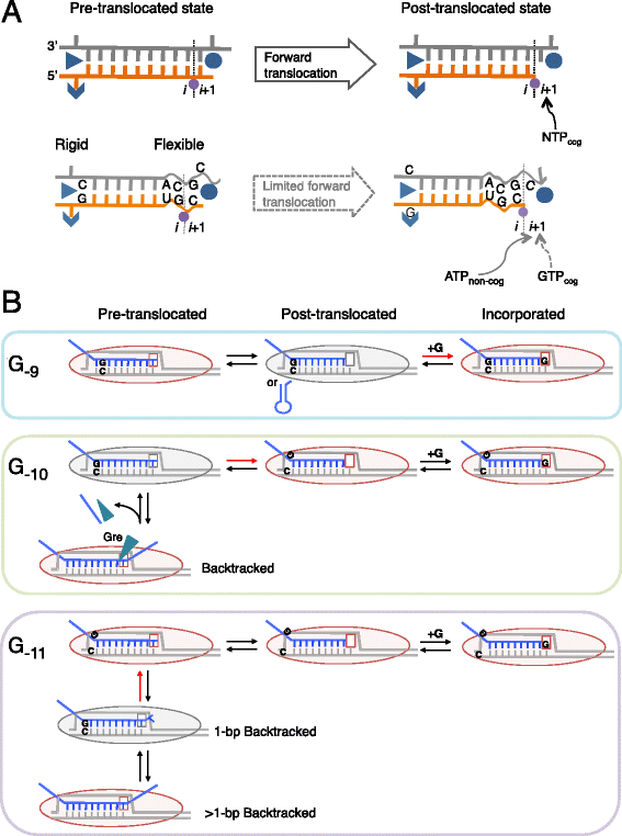
Structural and kinetic models of transcription pausing in vivo. (a) Structural model. RNAP elongation in a pause-free sequence (top) or the PIE (bottom) is shown. RNA (orange), template DNA strand (gray), catalytic Mg2+ (magenta), and two RNAP domains (blue) involved in the 5′ RNA separation from the RNA-DNA hybrid, i.e., Switch 3 (arrow head), lid (triangle) domain, and the bridge helix of RNAP (blue circle) are shown. The 3′ RNA-binding site (i) and the NTP binding site (i + 1) are also indicated. The 3′ ACGC 5′ sequence in the template DNA and the complementary 5′ UGC 3′ RNA sequence increase the flexibility of their backbones, which decreases cognate GTP (GTP cog) addition and increases non-cognate ATP (ATP non-cog) addition to the 3′ RNA end. The two RNAP domains can interact with riboG-dC at the upstream end of the hybrid, which interferes with the hybrid movement through the catalytic cleft of RNAP. (b) Kinetic model. RNAP pauses in the post-translocated (G−9 or RNA hairpin, top), pre-translocated (G−10, middle), and backtracked states (G−11, bottom). RNAPs with the i + 1 NTP binding site are shown (oval shapes with empty squares). Gre factors are indicated by cyan triangles. Post- and pre-translocated pauses were mainly observed in WT cells, and backtracked pauses were observed in ΔgreAB cells. The rate-determining steps during elongation are indicated by red arrows. The RNAP conformations captured by RNET-seq are indicated by gray ovals. Note that the GreAB-dependent cleavage, which occurs between i and i + 1 sites, ultimately converts the backtracked state to the post-translocated state. This state is rapidly converted back to the pre-translocated state prior to the next NTP binding and bond formation at the i + 1 site. The presence of activation energy much higher than k B T in each rate-determining step is assumed for the kinetic description of pausing in vivo [1, 39]

Similar articles

Cited by

References

    1. Imashimizu M, Shimamoto N, Oshima T, Kashlev M. Transcription elongation: Heterogeneous tracking of RNA polymerase and its biological implications. Transcription. 2014;5:e28285–28281–28210. doi: 10.4161/trns.28285. - DOI - PMC - PubMed
    1. Bailey MJ, Hughes C, Koronakis V. RfaH and the ops element, components of a novel system controlling bacterial transcription elongation. Mol Microbiol. 1997;26:845–51. doi: 10.1046/j.1365-2958.1997.6432014.x. - DOI - PubMed
    1. Tomar SK, Artsimovitch I. NusG-Spt5 proteins-Universal tools for transcription modification and communication. Chem Rev. 2013;113:8604–19. doi: 10.1021/cr400064k. - DOI - PMC - PubMed
    1. Adelman K, Lis JT. Promoter-proximal pausing of RNA polymerase II: emerging roles in metazoans. Nat Rev Genet. 2012;13:720–31. doi: 10.1038/nrg3293. - DOI - PMC - PubMed
    1. Perdue SA, Roberts JW. Sigma(70)-dependent transcription pausing in Escherichia coli. J Mol Biol. 2011;412:782–92. doi: 10.1016/j.jmb.2011.02.011. - DOI - PubMed

Publication types

MeSH terms

Associated data