

OCT angiography and sequential quantitative analysis of type 2 neovascularization after ranibizumab therapy
- PMID: 25976641
- PMCID: PMC4506353
- DOI: 10.1038/eye.2015.80
OCT angiography and sequential quantitative analysis of type 2 neovascularization after ranibizumab therapy
Abstract
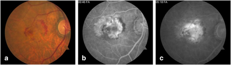
Purpose: To study the precise structural aspects of a type 2 neovascular membrane in a patient with age-related macular degeneration (AMD) using optical coherence tomography (OCT) angiography and perform sequential quantitative analysis of the membrane after ranibizumab therapy.
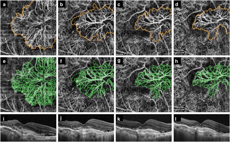
Patients and methods: Split-spectrum amplitude-decorrelation (SSADA) OCT angiography macular cubes (3 × 3 mm) were acquired with a light source centered at 840 nm, a bandwidth of 45 nm, and an A-scan-rate of 70 000 scans per second. Visible pathologic vessels were outlined manually on average intensity projection en face images, and the area of the lesion and the vessel density were measured at baseline and follow-up.
Results: At baseline, the neovascular lesion measured 4.12 mm(2) and the vessel density was 19.83 mm(-1). Four weeks after the first, and 2 and 4 weeks after the second ranibizumab injection, OCT angiography revealed a progressively smaller vascular lesion (2.32, 1.77 and 1.64 mm(2)), and vessel density (10.24, 8.52 and 7.57 mm(-1)), although the large central trunks of the lesion were unchanged.
Conclusions: In this study, an obvious reduction in size and vessel density of the neovascular lesion was noted after treatment with ranibizumab using SSADA OCT angiography technology. Microvascular components can be delineated with precision, suggesting that this technique may be useful for the management of patients with neovascular AMD in a clinical setting as well as for future clinical trials.
Figures
References
-
- Spaide RF, Klancnik Jr, JM, Cooney MJ. Retinal vascular layers imaged by fluorescein angiography and optical coherence tomography angiography. JAMA Ophthalmol. 2015;133:45–50. - PubMed
-
- Freund KB, Zweifel SA, Engelbert M. Do we need a new classification for choroidal neovascularization in age-related macular degeneration. Retina. 2010;30:1333–1349. - PubMed
Publication types
MeSH terms
Substances
LinkOut - more resources
Full Text Sources
Other Literature Sources
Research Materials
