. 2016 Jan;27(1):79-90.
doi: 10.1681/ASN.2014101030. Epub 2015 May 14.

Renal Production, Uptake, and Handling of Circulating αKlotho

Affiliations

Renal Production, Uptake, and Handling of Circulating αKlotho

Ming Chang Hu et al. J Am Soc Nephrol. 2016 Jan.

Abstract

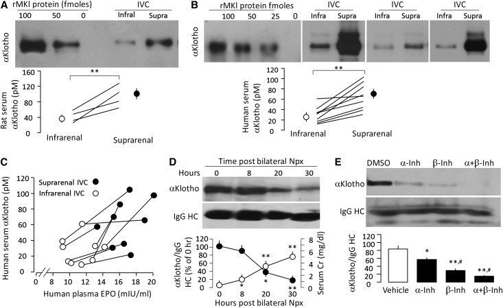
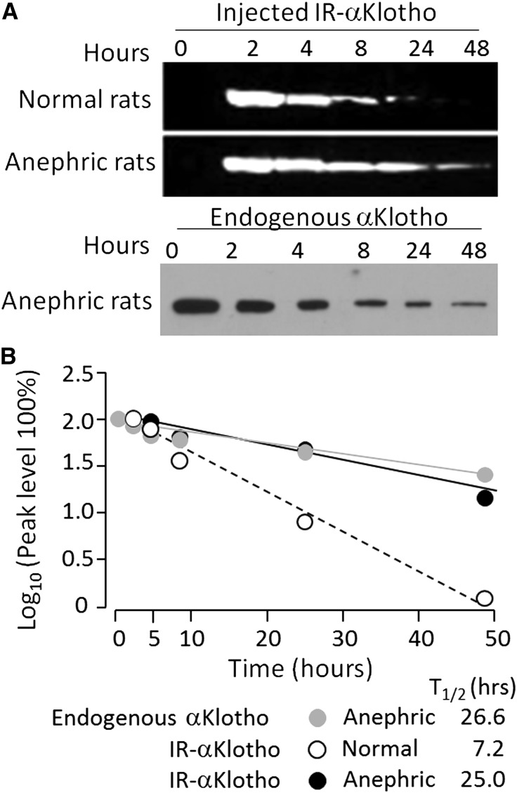
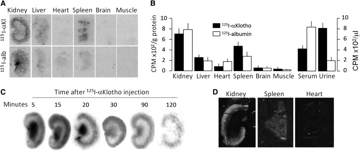
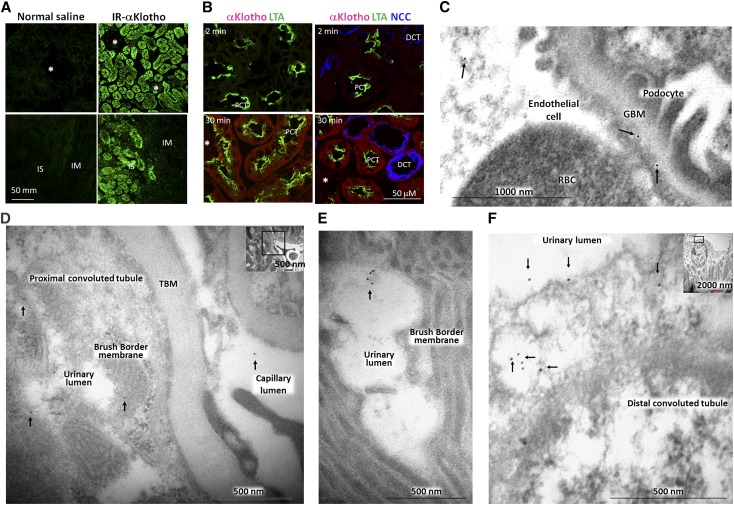
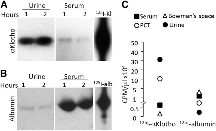
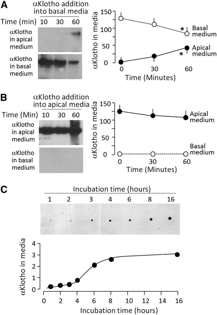
αKlotho is a multifunctional protein highly expressed in the kidney. Soluble αKlotho is released through cleavage of the extracellular domain from membrane αKlotho by secretases to function as an endocrine/paracrine substance. The role of the kidney in circulating αKlotho production and handling is incompletely understood, however. Here, we found higher αKlotho concentration in suprarenal compared with infrarenal inferior vena cava in both rats and humans. In rats, serum αKlotho concentration dropped precipitously after bilateral nephrectomy or upon treatment with inhibitors of αKlotho extracellular domain shedding. Furthermore, the serum half-life of exogenous αKlotho in anephric rats was four- to five-fold longer than that in normal rats, and exogenously injected labeled recombinant αKlotho was detected in the kidney and in urine of rats. Both in vivo (micropuncture) and in vitro (proximal tubule cell line) studies showed that αKlotho traffics from the basal to the apical side of the proximal tubule via transcytosis. Thus, we conclude that the kidney has dual roles in αKlotho homeostasis, producing and releasing αKlotho into the circulation and clearing αKlotho from the blood into the urinary lumen.

Keywords: Cell & Transport Physiology; Nephrectomy; Transcytosis; [REMOVED HYPERLINK FIELD]Klotho; distal tubule; renal proximal tubule cell.


Figures

Figure 1.
Figure 1.
The kidney contributes to circulating blood αKlotho. (A) αKlotho protein levels in inferior vena cava (IVC) in normal SD rats. Upper panel shows immunoblot of αKlotho protein immunoprecipitated with anti-αKlotho antibody from 0.1 ml serum from infrarenal and suprarenal IVC. Different amounts of recombinant full-length murine αKlotho protein (rMKl) in left three lanes. Bottom panel show four independent animals with each one connected by lines. Means±SD are shown as circles and error bars. Statistical significance was assessed by paired t test, and accepted when **P<0.01. (B) αKlotho protein levels in human IVC. Upper panel shows immunoblot of αKlotho proteins immunoprecipitated with anti-αKlotho antibody from 0.1 ml serum from infrarenal and suprarenal IVC from three subjects. Bottom panel is a summary from nine patients and significant difference between infrarenal and suprarenal IVC was assessed by paired t test, and significant differences were accepted when **P<0.01. (C) Serum αKlotho levels (y axis) were plotted against serum erythropoietin (EPO) levels (x axis) measured in the same sample. The infrarenal and suprarenal samples from the same individuals are connected by lines. (D) Change in endogenous serum αKlotho in anephric rats subjected to bilateral nephrectomy (Npx). Blood was drawn at the specified time points post-surgery. Serum αKlotho was determined by immunoprecipitation-immunoblot. Upper panel is a representative blot. Bottom panel is a summary of serum αKlotho and serum Cr from three independent experiments. Data are expressed as means±SD. Statistical significance was assessed by one-way ANOVA followed by Student–Newman–Keuls test, and significant differences were accepted when *P<0.05; **P<0.01* versus hour 0. (E) WT mice were injected intraperitoneally with α- or/and β-secretase inhibitors (α-Inh, β-Inh) for 2 days and serum αKlotho was determined by immunoprecipitation-immunoblot. Upper panel is one set of representative data. Bottom panel is a summary of arbitrary unit of serum αKlotho over IgG HC from three independent experiments. Statistical significance was assessed by one-way ANOVA followed by post hoc Student–Newman–Keuls test, and was considered significant when *P<0.05 or **P<0.01 versus vehicle (DMSO); #P<0.05 versus α-secretase inhibitor.
Figure 2.
Figure 2.
The kidney clears circulating αKlotho. (A) Anephric rats were prepared by bilateral nephrectomy (Npx). Exogenous recombinant αKlotho protein labeled with infrared dye (IR-αKlotho) was injected and blood was drawn at the specified time points post-surgery and labeled αKlotho in the blood is shown in the upper panel. Endogenous αKlotho in serum of rats after Npx is shown in the bottom panel. Three independent experiments were performed showing similar results. (B) Half-life (T1/2) of exogenous αKlotho in normal and anephric rats and of endogenous αKlotho protein in anephric rats.
Figure 3.
Figure 3.
Tissue distribution of exogenous recombinant mouse αKlotho in normal rats. 125I-αKlotho and 125I-albumin were intravenously injected into normal Sprague-Dawley rats and blood, urine, and organs were collected. (A) Representative autoradiographs of organs harvested from rats injected with 125I-αKlotho and 125I-albumin. (B) Radioactivity of 125I-αKlotho and 125I-albumin in organs normalized to protein concentration (left panel), and of 125I-αKlotho and 125I-albumin in serum and urine (right panel). Data summarized from four independent experiments. (C) 125I-labeled αKlotho protein was intravenously injected into normal rats and kidneys were harvested at specific time points and cryosections were made for autoradiography. (D) Infrared-labeled αKlotho was intraperitoneally injected into normal rats. Two hours later, kidney, spleen, and heart were harvested, sectioned, and scanned with IR imager system.
Figure 4.
Figure 4.
The distribution of exogenous recombinant mouse αKlotho in the kidney. (A) Infrared dye labeled αKlotho (IR-αKlotho) or normal saline was intraperitoneally injected into normal rats. Two hours later, kidney was harvested, sectioned, and imaged with IR microscope. (B) αKlotho labeled with two types of fluorescent dye: Alexa 555 C2 Maleimide (λex 555 nm, λem 565 nm) and TAMRA-SE (λex 546 nm, λex 579 nm) was intravenously injected into normal rats, and kidneys were collected at specific time points and subjected to immunofluorescent staining for αKlotho, NaCl cotransporter (NCC), and Lotus-Tetragonolobus lectins (LTA). Asterisks depict glomerulus. DCT: distal convoluted tubule; IM: inner medulla; IS: inner strip of outer medulla; PCT: proximal convoluted tubule. (C–F). αKlotho protein was intraperitoneally injected into homozygous αKlotho-deficient mice and the kidneys were harvested for immunogold electron microscopy. Representative electron micrographs of exogenous αKlotho in (C) glomeruli, (D, E) proximal tubules, (F) distal tubules. Arrows indicate gold particle (labeled αKlotho protein).
Figure 5.
Figure 5.
Intact exogenous αKlotho is present in blood and urine. 125I-αKlotho and 125I-albumin were intravenously injected into normal rats and blood and urine were collected 1 and 2 hours after injection, respectively. (A) 125I-αKlotho and (B) 125I-albumin in urine and serum were visualized on SDS-PAGE. (C) Urine from free-flow micropuncture originating from PCT, Bowman’s space, bladder urine, and serum of rats injected with 125I-αKlotho or 125I-albumin, were collected at 1 hour after injection and the same volumes were subjected to scintillation counting.
Figure 6.
Figure 6.
αKlotho transcytosis from basal to apical side in OK cells. OK cells were seeded on Transwell plates and grown to confluence to separate apical and basolateral side. (A) αKlotho with C-terminal 6His tag was added to the basal medium. At given time points after incubation, media were collected from both apical and basal side, and subjected to immunoblot to quantify exogenous αKlotho protein with anti-His antibody (left panel). Right panel is a summary of total amount of αKlotho at each time point in each compartment. Data are expressed as means±SD from three independent experiments. Statistical significance was assessed by one-way ANOVA followed by Student–Newman–Keuls test, and significant differences were accepted when *P<0.05 versus 0 min. (B) αKlotho with C-terminal 6His tag was added to the apical medium. At given time points after incubation, media were collected from apical and basal side and subjected to immunoblot for quantitation of αKlotho protein with anti-His antibody (left panel). Right panel is a summary of total amount of αKlotho at each time point in each compartment. Data are expressed as means±SD from three independent experiments. Statistical significance was assessed by one-way ANOVA followed by Student–Newman–Keuls test. None of the changes were statistically significant. (C). Time course of transcytosis. Media were collected from the apical side when αKlotho with C-terminal 6His tag was added to the basal side, and subjected to dot blot αKlotho protein with anti-His antibody (upper panel). Bottom panel shows arbitrary densitometric units of αKlotho protein. Three independent experiments showed identical results.

References

    1. Kuro-o M, Matsumura Y, Aizawa H, Kawaguchi H, Suga T, Utsugi T, Ohyama Y, Kurabayashi M, Kaname T, Kume E, Iwasaki H, Iida A, Shiraki-Iida T, Nishikawa S, Nagai R, Nabeshima YI: Mutation of the mouse Klotho gene leads to a syndrome resembling ageing. Nature 390: 45–51, 1997 - PubMed
    1. Kurosu H, Yamamoto M, Clark JD, Pastor JV, Nandi A, Gurnani P, McGuinness OP, Chikuda H, Yamaguchi M, Kawaguchi H, Shimomura I, Takayama Y, Herz J, Kahn CR, Rosenblatt KP, Kuro-o M: Suppression of aging in mice by the hormone Klotho. Science 309: 1829–1833, 2005 - PMC - PubMed
    1. Hu MC, Kuro-o M, Moe OW: Renal and extrarenal actions of Klotho. Semin Nephrol 33: 118–129, 2013 - PMC - PubMed
    1. Hu MC, Shiizaki K, Kuro-o M, Moe OW: Fibroblast growth factor 23 and Klotho: physiology and pathophysiology of an endocrine network of mineral metabolism. Annu Rev Physiol 75: 503–533, 2013 - PMC - PubMed
    1. Ito S, Kinoshita S, Shiraishi N, Nakagawa S, Sekine S, Fujimori T, Nabeshima YI: Molecular cloning and expression analyses of mouse βKlotho, which encodes a novel Klotho family protein. Mech Dev 98: 115–119, 2000. - PubMed

Publication types

LinkOut - more resources