. 2015 Jun 5;348(6239):1151-4.
doi: 10.1126/science.aaa9344. Epub 2015 May 14.

Neurodegeneration. C9ORF72 repeat expansions in mice cause TDP-43 pathology, neuronal loss, and behavioral deficits

Affiliations

Neurodegeneration. C9ORF72 repeat expansions in mice cause TDP-43 pathology, neuronal loss, and behavioral deficits

Jeannie Chew et al. Science. .

Abstract

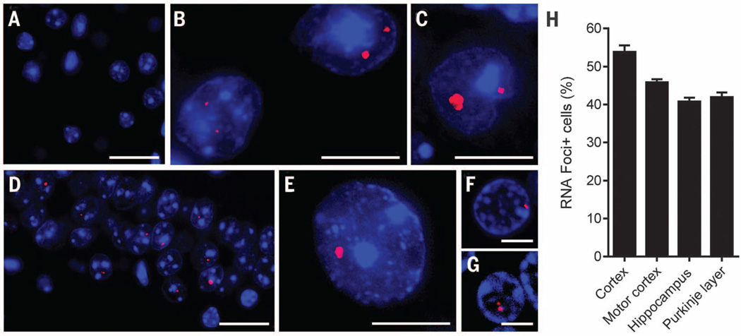
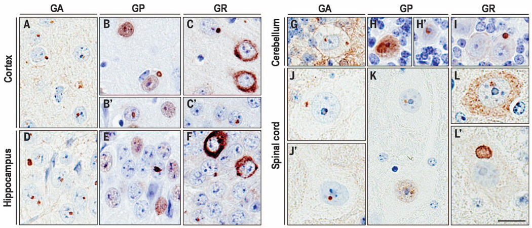
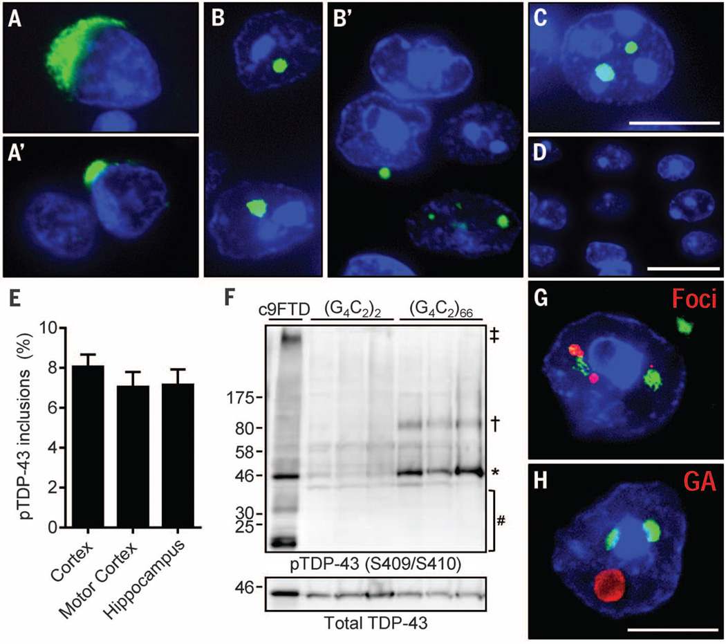
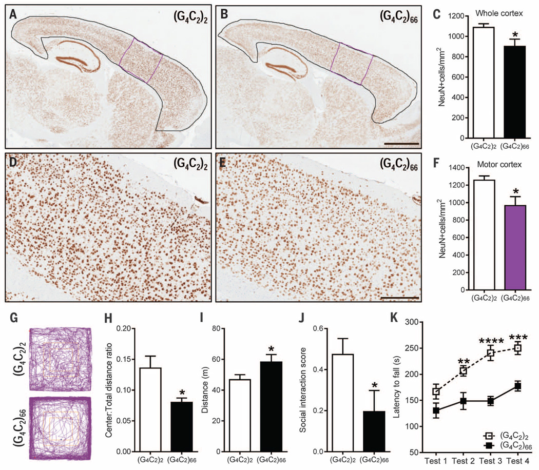
The major genetic cause of frontotemporal dementia and amyotrophic lateral sclerosis is a G4C2 repeat expansion in C9ORF72. Efforts to combat neurodegeneration associated with "c9FTD/ALS" are hindered by a lack of animal models recapitulating disease features. We developed a mouse model to mimic both neuropathological and clinical c9FTD/ALS phenotypes. We expressed (G4C2)66 throughout the murine central nervous system by means of somatic brain transgenesis mediated by adeno-associated virus. Brains of 6-month-old mice contained nuclear RNA foci, inclusions of poly(Gly-Pro), poly(Gly-Ala), and poly(Gly-Arg) dipeptide repeat proteins, as well as TDP-43 pathology. These mouse brains also exhibited cortical neuron and cerebellar Purkinje cell loss, astrogliosis, and decreased weight. (G4C2)66 mice also developed behavioral abnormalities similar to clinical symptoms of c9FTD/ALS patients, including hyperactivity, anxiety, antisocial behavior, and motor deficits.


Figures

Fig. 1
Fig. 1. Intranuclear RNA foci were detected in the CNS of (G4C2)66 mice
The presence of r(G4C2) RNA foci in brain and spinal cord of 6-month-old (G4C2)2 and (G4C2)66 mice was evaluated by RNA fluorescence in situ hybridization. (A) Foci were not detected in (G4C2)2 mice; shown is a representative image of the cortex. (B to G) In (G4C2)66 mice, RNA foci were detected in various regions—including the cortex (B), cerebellum (C), hippocampus (D), and spinal cord (E)—and were similar to foci observed in the cortex (F) and cerebellum (G) of c9FTD/ALS patients. (H) The average percentage of cells containing nuclear RNA foci in the indicated brain regions of (G4C2)66 mice (n = 11) was calculated. The total number of cells counted per region was 500 for cortex, motor cortex, and hippocampus (CA1 to CA3) and 100 for Purkinje cells. Scale bars: (A), (D), (F), and (G), 25 µm; (B), (C), and (E), 10 µm.
Fig. 2
Fig. 2. c9RAN protein pathology was detected throughout the CNS of (G4C2)66 mice
Intranuclear and cytoplasmic inclusions immunopositive for poly(GA), poly(GP), or poly(GR) were observed in various regions of the CNS of 6-month-old (G4C2)66 mice, including the cortex (A to C′), hippocampus (D to F), cerebellum (G to I), and spinal cord (J to L′). Note also the diffuse nuclear poly(GP) staining [(B), (B′), (E), (H), and (K)] and diffuse cytoplasmic poly(GR) staining [(C), (F), and (L)]. Semiquantitative analysis of c9RAN protein staining in different neuroanatomical regions is provided in table S1. No c9RAN proteins were detected in 6-month-old control (G4C2)2 mice (fig. S3). Scale bar, 20 µm.
Fig. 3
Fig. 3. (G4C2)66 mice developed inclusions composed of pTDP-43
(A and A′) Representative pTDP-43 inclusions in postmortem c9FTD/ALS frontal cortex sections. (B to D) Similarly, multiple cells with nuclear, and occasionally cytoplasmic pTDP-43, inclusions were observed in the cortex (B and B′) and hippocampus (C) of 6-month-old (G4C2)66 mice. No pTDP-43 immunoreactivity was detected in control (G4C2)2 mice; shown is a representative image of the cortex (D). (E) The average percentage of cells with pTDP-43 inclusions in the indicated regions was calculated on the basis of evaluations of 500 cells per mouse (n = 6 mice). (F) pTDP-43 was detected in brain urea fractions from (G4C2)66 mice and a c9FTD patient but not from (G4C2)2 mice. Symbols: ‡high molecular weight species; †oligomers; *full-length protein; #cleavage products. (G) Of 250 cells containing pTDP-43 inclusions examined among five (G4C2)66 mice, all were found to contain nuclear foci. (H) Of 250 cells containing pTDP-43 inclusions examined among five (G4C2)66 mice, ~75% were found to be positive for poly(GA) inclusions. Nuclei are stained blue in (A) to (D), (G), and (H). Scale bars: (A) to (C), (G), and (H), 10 µm; (D), 20 µm.
Fig. 4
Fig. 4. (G4C2)66 mice developed cortical neuron loss and exhibited behavior and motor deficits at 6 months of age
(A and B) Representative images of NeuN-labeled cells in the cortex of (G4C2)2 mice (n = 6) and (G4C2)66 mice (n = 6). (C) Quantification of NeuN-positive cells in the whole cortex [area delineated in black in (A) and (B)]. (D and E) Enhanced magnification of the motor cortex [area delineated in purple in (A) and (B)]. (F) Quantification of NeuN-positive cells in the motor cortex. (G to I) In the open-field test, (G4C2)66 mice (n = 11) displayed signs of increased anxiety-like behavior compared with (G4C2)2 mice (n = 12), as evidenced by representative traces (G) and a significant decrease in distance traveled in the center area normalized to total distance traveled (H). (G4C2)66 mice traveled a greater distance overall (I), which is indicative of hyperactivity. (J) In the social interaction test, (G4C2)66 mice had a decreased interaction score, which is indicative of social deficits. (K) Motor deficits, manifesting as a decreased latency to fall from a rotating rod, were observed in (G4C2)66 mice on test day 2 to 4 of the Rota-Rod test. Scale bars: (A) and (B), 1 mm; (D) and (E), 300 µm. Data are presented as means T SEM; *P < 0.05, unpaired, two-tailed Student’s t-test. ****P < 0.0001, ***P < 0.001, **P < 0.01, two-way analysis of variance followed by Sidak’s multiple comparisons test. Abbreviated units: m, meters; s, seconds.

Comment in

References

    1. DeJesus-Hernandez M, et al. Neuron. 2011;72:245–256. - PMC - PubMed
    1. Renton AE, et al. Neuron. 2011;72:257–268. - PMC - PubMed
    1. Mizielinska S, et al. Science. 2014;345:1192–1194. - PMC - PubMed
    1. Zhang YJ, et al. Acta Neuropathol. 2014;128:505–524. - PMC - PubMed
    1. May S, et al. Acta Neuropathol. 2014;128:485–503. - PMC - PubMed

Publication types

MeSH terms