. 2015 May 15;10(5):e0126504.
doi: 10.1371/journal.pone.0126504. eCollection 2015.

Modes of Escherichia coli Dps Interaction with DNA as Revealed by Atomic Force Microscopy

Affiliations

Modes of Escherichia coli Dps Interaction with DNA as Revealed by Atomic Force Microscopy

Vladislav V Melekhov et al. PLoS One. .

Abstract

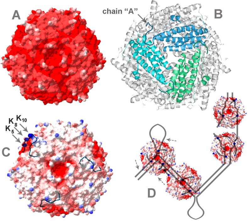
Multifunctional protein Dps plays an important role in iron assimilation and a crucial role in bacterial genome packaging. Its monomers form dodecameric spherical particles accumulating ~400 molecules of oxidized iron ions within the protein cavity and applying a flexible N-terminal ends of each subunit for interaction with DNA. Deposition of iron is a well-studied process by which cells remove toxic Fe2+ ions from the genetic material and store them in an easily accessible form. However, the mode of interaction with linear DNA remained mysterious and binary complexes with Dps have not been characterized so far. It is widely believed that Dps binds DNA without any sequence or structural preferences but several lines of evidence have demonstrated its ability to differentiate gene expression, which assumes certain specificity. Here we show that Dps has a different affinity for the two DNA fragments taken from the dps gene regulatory region. We found by atomic force microscopy that Dps predominantly occupies thermodynamically unstable ends of linear double-stranded DNA fragments and has high affinity to the central part of the branched DNA molecule self-assembled from three single-stranded oligonucleotides. It was proposed that Dps prefers binding to those regions in DNA that provide more contact pads for the triad of its DNA-binding bundle associated with one vertex of the protein globule. To our knowledge, this is the first study revealed the nucleoid protein with an affinity to branched DNA typical for genomic regions with direct and inverted repeats. As a ubiquitous feature of bacterial and eukaryotic genomes, such structural elements should be of particular care, but the protein system evolutionarily adapted for this function is not yet known, and we suggest Dps as a putative component of this system.


Conflict of interest statement

Competing Interests: The authors have declared that no competing interests exist.

Figures

Fig 1
Fig 1. [A]: An example of electrophoretic mobility shift assays performed as described in Matherials and Methods.
One pmol of the 214 bp-long DNA fragment amplified with primers dps_F1 and dps_R1 (designated as F1-R1) was loaded on the lane 1 alone as independent marker. Other samples contained two fragments (1 pmol each) and Dps as indicated above the photo. The gel was calibrated by marker ladder (M). [B] DNAse I footprinting assays performed for Dps complexes with F1-R1, F2-R2 and F1-R2 DNA fragments. 32P-labeled primers are indicated by asterisks. Gels were calibrated by the products of G-sequencing ladder. Positional marks show distance to 32P-labeled primers F1 or R2. Protected sites are dashed. Hyperreactive sites are not marked. [C]: Scheme illustrating relative disposition and structural organization of analyzed fragments. Direct and inverted repeats in their sequences are enhanced and additionally indicated by arrows. Corresponding bands in [B] are denoted by open and gray arrows for direct and inverted repeats, respectively. Bent arrow points transcription start site in Pdps promoter.
Fig 2
Fig 2. AFM images.
[A]: Dps protein; [B-D]: DNA-fragments containing correspondingly complete regulatory region of gene dps (420 bp, primers dps_F1 and dps_R2), its proximal (259 bp, primers dps_F2 and dps_R2) and distal (214 bp, primers dps_F1 and dps_R1) part. Panels b and c: complexes formed by Dps with 214 bp (b) and 259 bp (c) DNA-fragments. White bar scales represent 100 nm.
Fig 3
Fig 3. Complexes formed by Dps with four artificial branched constructs schematically drown on each section.
The sequence of colored circles corresponds to the sequence of the oligonucleotides used (Table 1). Panels demonstrate AFM images obtained for free DNA samples and their complexes with Dps (left and right scans, respectively). Assembling of DNA constructs and complex formation were performed as described in Materials and Methods. Insert in the right panel of Fig D exemplifies the 3D image of complexes formed with Y5_Y6_Y8 triplex. Ends of all three branches are clearly visible. White bars represent 100 nm scales.
Fig 4
Fig 4. Electrophoretic mobility shift assays performed for Dps complexes with Y-shape constructs Y1_Y2_Y3 (drown in Fig 1B) and Y5_Y6_Y7 (similar to Y5_Y6_Y8 drown in Fig 1D but without single-stranded loop).
The composition of the samples and the molar Dps:DNA ratio are indicated above photos. Branched DNA molecules were assembled from indicated oligonucleotides mixed in equal concentration (2–5 pmol each) and prepared as described in Materials and Methods. Without prior fractionation they were used for complex formation with Dps. The amount of Dps was chosen based on the assumption that all the oligonucleotides formed triplex, as in the case of Y5_Y6_Y7. Indicated molar ratios for Y1_Y2_Y3 are, therefore, overestimated. Gels were calibrated by marker DNA fragments (M) and stained by AgNO3 so as to visualize both DNA molecules and Dps.
Fig 5
Fig 5. Examples of native (A, C) and nicked (B, D, E) plasmids pET28b in free state (A, B) and forming complexes with Dps (C-E).
White bars scale images (nm). Horizontal and vertical arrows in panels C-E point out complexes with lower and higher levels of oligomerization, respectively.
Fig 6
Fig 6. Crystal structure of Dps (PDB ID: 1DPS) was obtained with 1.6Å resolution by Grant et al [16].
Charge distribution on the surface of Dps was calculated using the Swiss-PdbViewer version 4.1.0 [52]. In panel [A] the threshold for Coulombic surface coloring was set at -12 for red (negative electrostatic potential), at -1.5 for white and at 0 for blue (positive potential). In panel [C] the scale was changed to: -12, -4.8 and 0, respectively. N-terminus of chain “A” was proportionally lengthened so as to show schematically the location of Lys5, and Lys8 (blue balls). N-termini of two other chains were lengthened up to position 9 (as in chain “A”). Gray asterisks in [C] mark ends of the flexible modules. Panel [B] shows the central pore and the disposition of subunits near the same vertex. Panel [D] schematically illustrates different modes of interaction, including binding to branched DNA of slipped loop structure (left), nicked DNA (two right particles), and straight DNA (second from the right). In the latter case, two N-termini are involved in DNA binding, while the third one of the same vertex can bind the vacant site of the negatively charged spot on the surface of the other Dps molecule. Even though such protein-protein contacts may be formed in solution, the presence of DNA must stabilize them and promote the protein aggregation. All molecular dimensions on the scheme are set in proportion to natural sizes.

Similar articles

Cited by

References

    1. Azam TA, Ishihama A. Twelve species of the nucleoid-associated protein from Escherichia coli. Sequence recognition specificity and DNA binding affinity. J Biol Chem. 1999;274: 33105–33113. 10.1074/jbc.274.46.33105 - DOI - PubMed
    1. Dorman CJ. Nucleoid-associated proteins and bacterial physiology. Adv Appl Microb. 2009;67: 47–64. 10.1016/S0065-2164(08)01002-2 - DOI - PubMed
    1. Azam TA, Iwata A, Nishimura A, Ueda S, Ishihama A. Growth phase-dependent variation in protein composition of the Escherichia coli nucleoid. J Bacteriol. 1999;181: 6361–6370. - PMC - PubMed
    1. Goodman SD, Velten NJ, Gao Q, Robinson S, Segall AM. In vitro selection of integration host factor binding sites. J Bacteriol. 1999;181: 3246–3255. - PMC - PubMed
    1. Cho B-K, Knight EM, Barrett CL, Palsson BO. Genome-wide analysis of Fis binding in Escherichia coli indicates a causative role for A-/AT-tracts. Gen Res. 2008;18: 900–910. 10.1101/gr.070276.107 - DOI - PMC - PubMed

Publication types

MeSH terms