. 2015 May 15;10(5):e0127475.
doi: 10.1371/journal.pone.0127475. eCollection 2015.

The Novel Gene CRNDE Encodes a Nuclear Peptide (CRNDEP) Which Is Overexpressed in Highly Proliferating Tissues

Affiliations

The Novel Gene CRNDE Encodes a Nuclear Peptide (CRNDEP) Which Is Overexpressed in Highly Proliferating Tissues

Lukasz Michal Szafron et al. PLoS One. .

Abstract

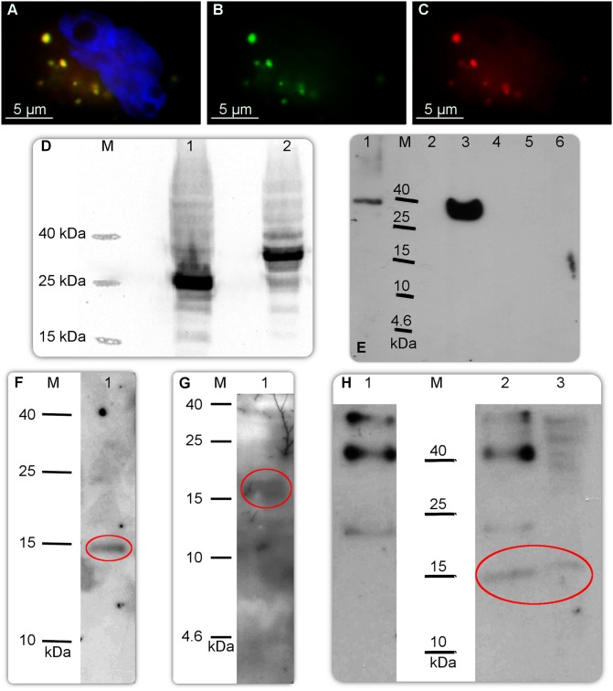
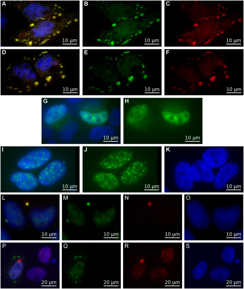
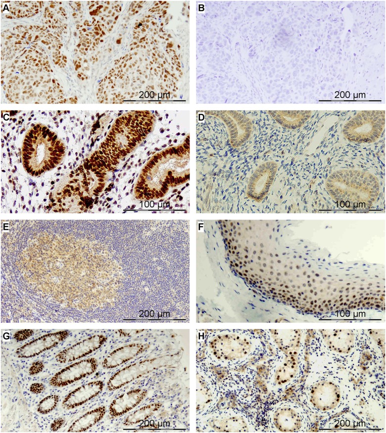
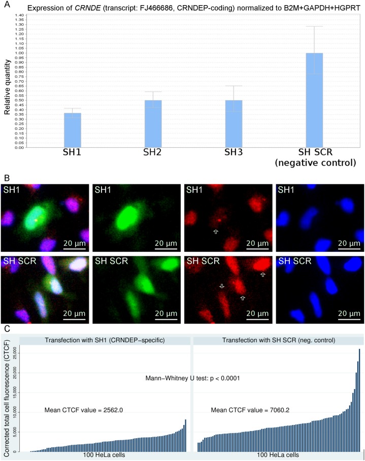
CRNDE, recently described as the lncRNA-coding gene, is overexpressed at RNA level in human malignancies. Its role in gametogenesis, cellular differentiation and pluripotency has been suggested as well. Herein, we aimed to verify our hypothesis that the CRNDE gene may encode a protein product, CRNDEP. By using bioinformatics methods, we identified the 84-amino acid ORF encoded by one of two CRNDE transcripts, previously described by our research team. This ORF was cloned into two expression vectors, subsequently utilized in localization studies in HeLa cells. We also developed a polyclonal antibody against CRNDEP. Its specificity was confirmed in immunohistochemical, cellular localization, Western blot and immunoprecipitation experiments, as well as by showing a statistically significant decrease of endogenous CRNDEP expression in the cells with transient shRNA-mediated knockdown of CRNDE. Endogenous CRNDEP localizes predominantly to the nucleus and its expression seems to be elevated in highly proliferating tissues, like the parabasal layer of the squamous epithelium, intestinal crypts or spermatocytes. After its artificial overexpression in HeLa cells, in a fusion with either the EGFP or DsRed Monomer fluorescent tag, CRNDEP seems to stimulate the formation of stress granules and localize to them. Although the exact role of CRNDEP is unknown, our preliminary results suggest that it may be involved in the regulation of the cell proliferation. Possibly, CRNDEP also participates in oxygen metabolism, considering our in silico results, and the correlation between its enforced overexpression and the formation of stress granules. This is the first report showing the existence of a peptide encoded by the CRNDE gene.


Conflict of interest statement

Competing Interests: The authors have declared that no competing interests exist.

Figures

Fig 1
Fig 1. In silico studies on CRNDE transcripts and the 84aas protein product, CRNDEP.
A) A graphical alignment of two complete CRNDE transcripts, previously identified by our research team (FJ466686, FJ466685) to four reference RNA sequences (NR_034105, NR_034106, NR_110453, NR_110454) available in GenBank. Our sequences are the most similar to the NR_034106 reference sequence, except for their exons 1b and 6, which are shorter. In addition, exon 5 in the FJ466686 transcript is shortened compared to the other transcripts. This allows for the formation of an alternative 84aas open reading frame (ORF), specific to this particular splice variant. ORFs are shown as yellow arrows, whereas pink arrows represent three epitopes used herein for development of the anti-CRNDEP antibody. B) Structures of three different fusion proteins used in this study after their enforced overexpression in HeLa cells: I—2xFLAG-CRNDEP (120 aas, 14.0 kDa), II—6xHis-CRNDEP-EGFP (346 aas, 39.2 kDa), III—DsRed Monomer-6xHis-CRNDEP (340 aas, 38.5 kDa). C) An alignment of several protein sequences obtained by using the 84aas CRNDEP sequence as a query to translated BLAST (NCBI tblastn). This bioinformatics tool searched the translated nucleotide collection (nr/nt) from all organisms and found hypothetical proteins highly similar to human CRNDEP. Interestingly, all the hits were confined to the Primates order only. D) An alignment between CRNDEP and the N-terminal consensus sequence of RNRs, generated by the Clustal Omega algorithm, showing a significant level of homology and conservation.
Fig 2
Fig 2. Molecular studies on CRNDEP.
A) Simultaneous overexpression of the 6xHis-CRNDEP-EGFP and DsRed Monomer-6xHis-CRNDEP fusion proteins in HeLa cells, visualized under a fluorescence microscope. The former protein glows green and the latter glows red in these conditions. Yellow glow is caused by a co-localization of these two fusion proteins. Nuclei were stained blue with DAPI. The same shot with only the green (B) or the red (C) channel shown. D) Western blot-based verification of the size of the 6xHis-CRNDEP-EGFP fusion protein. M—Spectra Multicolor Low Range Protein Ladder (Thermo-Fisher Scientific), 1—the EGFP reporter protein (26.9 kDa), 2—6xHis-CRNDEP-EGFP (39.2 kDa). E) Western blot-based verification of the specificity of our custom-made polyclonal anti-CRNDEP antibody. M—Spectra Multicolor Low Range Protein Ladder; 1—DsRed Monomer-6xHis-CRNDEP (340 aas, 38.5 kDa); 2—purified 14 kDa protein containing the 6xHis tag, 1.4 μg (a negative control of the antibody's specificity, non-commercial); 3—6xHis-CRNDEP-EGFP (346 aas, 39.2 kDa); 4—empty; 5—EGFP (239 aas, 26.9 kDa, a negative control); 6—DsRed Monomer (232 aas, 26.2 kDa, a negative control). A loading control (the PVDF membrane used in this experiment, stained with Ponceau S) is shown in S13 Fig. F–G) Detection of the overexpressed 2xFLAG-CRNDEP protein (~14 kDa) in a total protein lysate from 0.25 million HeLa cells with either the anti-FLAG (F) or anti-CRNDEP (G) antibody. H) Immunoprecipitation of 2xFLAG-CRNDEP using the anti-CRNDEP antibody (2) and control IgG (1) (both from a rabbit). A total protein lysate before immunoprecipitation was loaded for comparison (3). After precipitation, the 2xFLAG-CRNDEP protein was detected on the PVDF membrane using the anti-FLAG antibody. The correct bands in Fig F–H are encircled.
Fig 3
Fig 3. Cellular localization experiments.
A–H) Stress granules—localization studies of CRNDEP in a fusion with a fluorescent tag. The EYFP-TIA-1 fusion protein (specific to stress granules) glowed green, the DsRed Monomer-6xHis-CRNDEP fusion protein glowed red and nuclei were stained blue with DAPI. HeLa cells after co-transfection with both the pDsRed Monomer-C1_CRNDEP and pEYFP-TIA-1 plasmids (not treated with sodium arsenite) (A); the same shot with only the green (B) or red (C) channel shown. When this co-transfection was followed by the treatment with sodium arsenite (D), stress granules appeared as well; the same shot with only the green (E) or red (F) channel shown. On the contrary, a transfection of HeLa cells with the pEYFP-TIA-1 plasmid only (G) did not cause the formation of stress granules; the same shot with only the green channel shown (H). I–K) The results of immunofluorescence studies in HeLa cells. Endogenous CRNDEP was immunostained green using the custom-made primary rabbit anti-CRNDEP antibody in combination with the secondary fluorescein (FITC)-conjugated anti-rabbit antibody (I); the same shot with only the green channel shown (J). In order to check the reaction specificity, the cells were incubated with the secondary antibody only. The lack of a green glow is a proof that the staining is specific (K). L–O) HeLa cells after overexpression of the fusion protein DsRed Monomer-CRNDEP, not treated with sodium arsenite. This fusion protein glowed red under a fluorescence microscope, localized to stress granules and was absent in the nucleus (N). By contrast, endogenous CRNDEP (stained green with FITC) was present predominantly in the nucleus, where it formed grains (M). Overlapping of green and red signals in stress granules (L) proved that the anti-CRNDEP antibody is capable of specifically detecting both the endogenous and the artificially overexpressed variants of CRNDEP. P–S) HeLa cells transfected with the pYFP-TIA-1 plasmid and also treated with sodium arsenite. Stress granules were visible as green dots outside the nucleus, which was stained blue with DAPI. No co-localization between endogenous CRNDEP (red) and TIA-1 (green) was observed (P); the same shot with only the green (Q), red (R) and blue (S) channel shown.
Fig 4
Fig 4. The endogenous CRNDEP peptide expression in different human tissues evaluated by immunohistochemical stainings.
A) Epithelial ovarian cancer (serous carcinoma) with heterogeneous nuclear expression; B) The same section of ovarian carcinoma incubated with a blocking peptide; C) Normal proliferative phase endometrium with strong nuclear expression within the glandular epithelium and heterogeneous staining in stromal cells; D) Atrophic endometrium with negative staining in the nuclei; Normal tonsil (E-F) with heterogeneous (weak to moderate) nuclear expression in the germinal center (E) and strong nuclear expression in the parabasal layer of the squamous epithelium (F); G) Normal intestine: strong nuclear expression in intestinal crypts; H) Seminiferous tubules of an atrophic human testis: strong nuclear expression in spermatocytes.
Fig 5
Fig 5. Evaluation of shRNA-mediated knockdown of CRNDE.
The effects of CRNDE gene silencing were evaluated at either mRNA (A) or protein level (B-C). The strongest decrease in the amount of the CRNDEP-coding transcript (by ~65%) was observed for the SH1 silencing construct (A). The effects of this knockdown were detectable at the protein level as well (B, C), leading to a statistically significant decline in the amount of CRNDEP (red signal) in the cells transfected with the silencing construct (green signal). As expected, such a correlation did not occur in the cells transfected with the construct encoding a control (scrambled) shRNA molecule (SH SCR). The transfected cells are marked with white arrows.

References

    1. Cabili MN, Trapnell C, Goff L, Koziol M, Tazon-Vega B, Regev A, et al. Integrative annotation of human large intergenic noncoding RNAs reveals global properties and specific subclasses. Genes Dev. 2011;25: 1915–1927. 10.1101/gad.17446611 - DOI - PMC - PubMed
    1. Graham LD, Pedersen SK, Brown GS, Ho T, Kassir Z, Moynihan AT, et al. Colorectal Neoplasia Differentially Expressed (CRNDE), a Novel Gene with Elevated Expression in Colorectal Adenomas and Adenocarcinomas. Genes Cancer. 2011;2: 829–840. 10.1177/1947601911431081 - DOI - PMC - PubMed
    1. Zhang X, Sun S, Pu JKS, Tsang ACO, Lee D, Man VOY, et al. Long non-coding RNA expression profiles predict clinical phenotypes in glioma. Neurobiol Dis. 2012;48: 1–8. 10.1016/j.nbd.2012.06.004 - DOI - PubMed
    1. Szafron L, Lisowska K, Rubel T, Sobiczewski P, Kupryjanczyk J. ING1, PTPN2, PCID2, VGLL1 and LOC388279 genes as potential prognostic markers in ovarian cancer. Int J Mol Med. 2009;24: S40.
    1. Szafron L, Balcerak A, Grzybowska EA, Rubel T, Kupryjanczyk J. The CRNDE, VAV2 and CEBPA genes as new negative prognostic factors in ovarian cancer. Eur. J. Cancer. 2013;49: S215.

Publication types

LinkOut - more resources