. 2015 May 15;10(5):e0126688.
doi: 10.1371/journal.pone.0126688. eCollection 2015.

Loss of Serglycin Promotes Primary Tumor Growth and Vessel Functionality in the RIP1-Tag2 Mouse Model for Spontaneous Insulinoma Formation

Affiliations

Loss of Serglycin Promotes Primary Tumor Growth and Vessel Functionality in the RIP1-Tag2 Mouse Model for Spontaneous Insulinoma Formation

Andrew Hamilton et al. PLoS One. .

Abstract

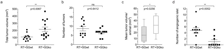
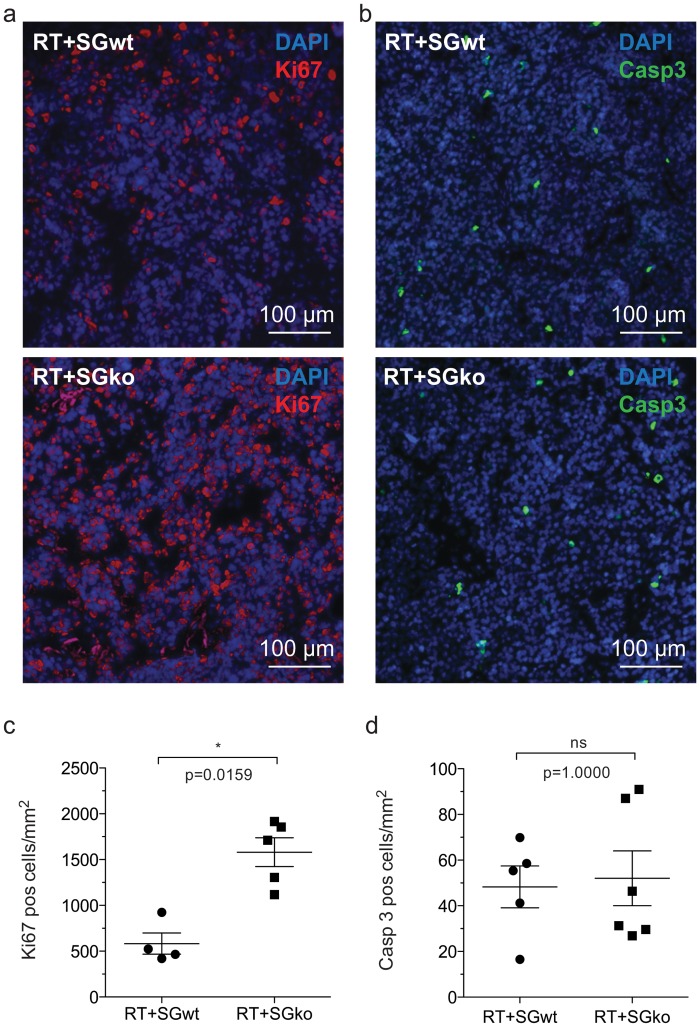
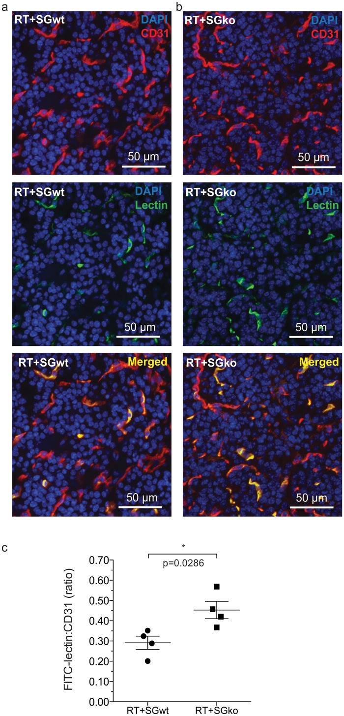
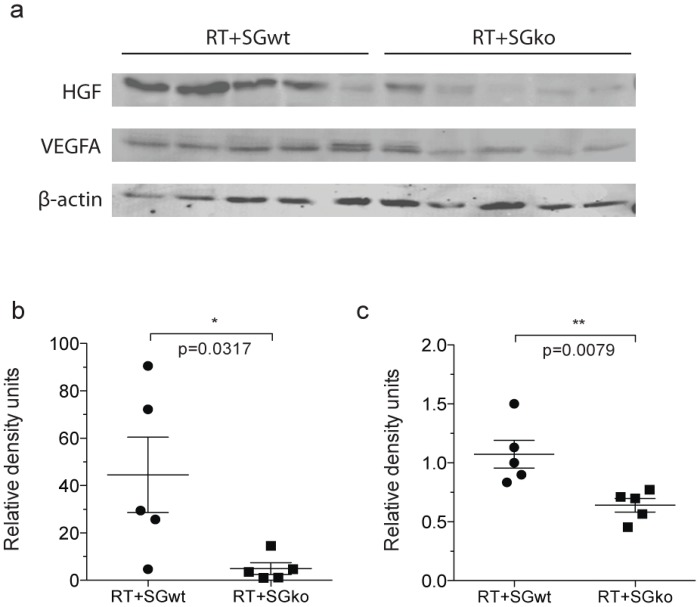
The serglycin proteoglycan is mainly expressed by hematopoietic cells where the major function is to retain the content of storage granules and vesicles. In recent years, expression of serglycin has also been found in different forms of human malignancies and a high serglycin expression level has been correlated with a more migratory and invasive phenotype in the case of breast cancer and nasopharyngeal carcinoma. Serglycin has also been implicated in the development of the tumor vasculature in multiple myeloma and hepatocellular carcinoma where reduced expression of serglycin was correlated with a less extensive vasculature. To further investigate the contribution of serglycin to tumor development, we have used the immunocompetent RIP1-Tag2 mouse model of spontaneous insulinoma formation crossed into serglycin deficient mice. For the first time we show that serglycin-deficiency affects orthotopic primary tumor growth and tumor vascular functionality of late stage carcinomas. RIP1-Tag2 mice that lack serglycin develop larger tumors with a higher proliferative activity but unaltered apoptosis compared to normal RIP1-Tag2 mice. The absence of serglycin also enhances the tumor vessel functionality, which is better perfused than in tumors from serglycin wild type mice. The presence of the pro-angiogenic modulators vascular endothelial growth factor and hepatocyte growth factor were decreased in the serglycin deficient mice which suggests a less pro-angiogenic environment in the tumors of these animals. Taken together, we conclude that serglycin affects multiple aspects of spontaneous tumor formation, which strengthens the theory that serglycin acts as an important mediator in the formation and progression of tumors.


Conflict of interest statement

Competing Interests: The authors have declared that no competing interests exist.

Figures

Fig 1
Fig 1. Tumor burden and number of angiogenic islets.
Loss of serglycin increases total and mean tumor volume, and decreases the number of angiogenic islets in RIP1-Tag2 mice. Tumors and angiogenic islets were excised from RTposSGwt and RTposSGko mice at 15 weeks of age and measured. Individual tumor volumes were calculated using the formula (π/6)×width 2 ×length, which was then used to determine total tumor volume (a). The frequency tumors (b) was also measured and used to calculate mean tumor volume per mouse (c). The number of angiogenic islets was also counted (d). Each data point in represents an individual animal. Statistical analysis was performed using a two-tailed Mann-Whitney test. Error bars represent mean ± SEM.
Fig 2
Fig 2. Proliferative and apoptotic status of tumor tissue.
The proliferative activity is increased in tumors from RTposSGko mice compared to those from RTposSGwt mice. Tumor sections from RTposSGwt and RTposSGko mice were stained for apoptotic and proliferative activity using antibodies against Ki67 (a) and cleaved caspase-3 (Casp-3, b) respectively. Positive cells were counted and tumors from serglycin deficient animals had a significantly increased levels of proliferation (c) while no difference was detected in apoptosis between the two genotypes. Statistical analysis was performed using a two-tailed Mann-Whitney test. Error bars represent mean ± SEM.
Fig 3
Fig 3. Tumor vessel perfusion.
The vascular function is increased in tumors of RTposSGko versus RTposSGwt mice. 15 week RTposSGwt (a) and RTposSGko (b) mice were perfused with FITC-lectin, which was introduced to the circulation prior to sacrifice of the animals and excision of the pancreas. Tumor sections were stained for the endothelial marker CD31 (red), the localization of which was compared to lectin (green) that had immobilized on the luminal side of endothelial cells in perfused vessels. The ratio between immobilized lectin and CD31 staining was calculated to determine the fraction of perfused, and thus functional vessels (c). Each data point in represents an individual animal. Statistical analysis was performed using a two-tailed Mann-Whitney test. Error bars represent mean ± SEM.
Fig 4
Fig 4. Protein levels of HFG and VEGF in tumor tissue.
The levels of HGF and VEGF are significantly decreased in the tumors of serglycin deficient RIP1-Tag2 mice. Lysed tumor material from 15w RTposSGwt and RTposSGko mice was probed for HGF and VEGF by western blotting (a). HGF (b) and VEGF (c) were significantly less abundant in the serglycin deficient tumors. Values were normalized to beta-actin (a). Each data point in represents an individual animal. Statistical analysis was performed using a two-tailed Mann-Whitney test. Error bars represent mean ± SEM.
Fig 5
Fig 5. Presence of inflammatory cells in tumor tissue.
Macrophage and neutrophil infiltration are unaffected by loss of serglycin in RIP1-Tag2 tumors. Tumor sections from 15w RTposSGwt and RTposSGko mice were immunostained from the neutrophil and macrophage markers Gr-1 (a) and F4/80 (b) respectively. For Gr-1, the number of positive cells/mm2 was calculated (c) and there was no difference in infiltration of neutrophils between the two groups. For F4/80, the area of positive staining was measured (d) and although there was a slight trend to decreased macrophage infiltration in serglycin deficient animals, this was not significant. Each data point in represents an individual animal. Statistical analysis was performed using a two-tailed Mann-Whitney test. Error bars represent mean ± SEM.

References

    1. Kolset SO, Pejler G. Serglycin: a structural and functional chameleon with wide impact on immune cells. J Immunol. 2011;187(10):4927–33. 10.4049/jimmunol.1100806 . - DOI - PubMed
    1. Schick BP, Gradowski JF, San Antonio JD. Synthesis, secretion, and subcellular localization of serglycin proteoglycan in human endothelial cells. Blood. 2001;97(2):449–58. Epub 2001/01/12. . - PubMed
    1. He L, Zhou X, Qu C, Tang Y, Zhang Q, Hong J. Serglycin (SRGN) overexpression predicts poor prognosis in hepatocellular carcinoma patients. Med Oncol. 2013;30(4):707 Epub 2013/09/03. 10.1007/s12032-013-0707-4 . - DOI - PubMed
    1. Korpetinou A, Skandalis SS, Moustakas A, Happonen KE, Tveit H, Prydz K, et al. Serglycin is implicated in the promotion of aggressive phenotype of breast cancer cells. PLoS One. 2013;8(10):e78157 Epub 2013/11/10. doi: 10.1371/journal.pone.0078157 PONE-D-13-33958 [pii]. - DOI - PMC - PubMed
    1. Li XJ, Ong CK, Cao Y, Xiang YQ, Shao JY, Ooi A, et al. Serglycin is a theranostic target in nasopharyngeal carcinoma that promotes metastasis. Cancer Res. 2011;71(8):3162–72. Epub 2011/02/04. doi: 10.1158/0008-5472.CAN-10-3557 0008-5472.CAN-10-3557 [pii]. . - DOI - PubMed

Publication types

MeSH terms