

Longitudinal changes in gestational weight gain and the association with intrauterine fetal growth
- PMID: 25978857
- PMCID: PMC4458401
- DOI: 10.1016/j.ejogrb.2015.04.006
Longitudinal changes in gestational weight gain and the association with intrauterine fetal growth
Abstract
Objective: Total pregnancy weight gain has been associated with infant birthweight; however, most prior studies lacked repeat ultrasound measurements. Understanding of the longitudinal changes in maternal weight gain and intrauterine changes in fetal anthropometrics is limited.
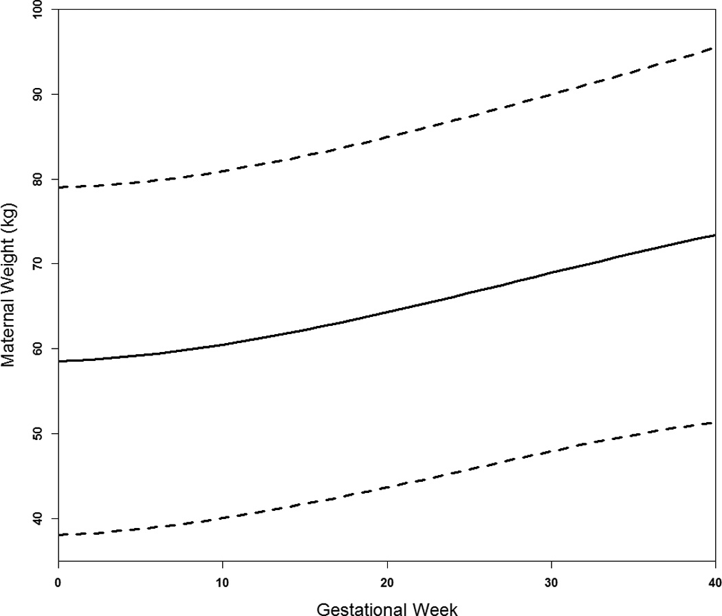
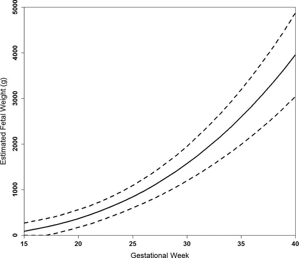
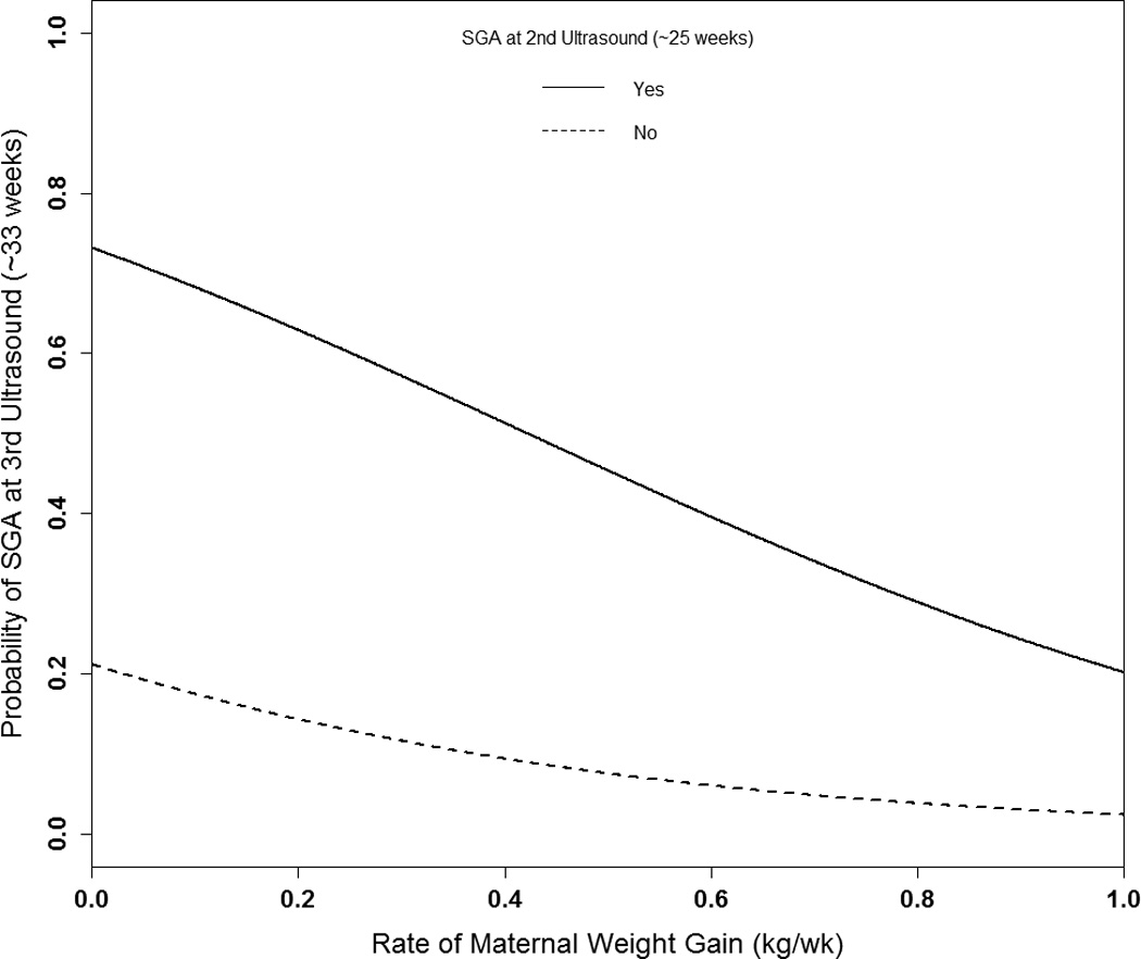
Study design: Prospective data from 1314 Scandinavian singleton pregnancies at high-risk for delivering small-for-gestational-age (SGA) were analyzed. Women had ≥1 (median 12) antenatal weight measurements. Ultrasounds were targeted at 17, 25, 33, and 37 weeks of gestation. Analyses involved a multi-step process. First, trajectories were estimated across gestation for maternal weight gain and fetal biometrics [abdominal circumference (AC, mm), biparietal diameter (BPD, mm), femur length (FL, mm), and estimated fetal weight (EFW, g)] using linear mixed models. Second, the association between maternal weight changes (per 5 kg) and corresponding fetal growth from 0 to 17, 17 to 28, and 28 to 37 weeks was estimated for each fetal parameter adjusting for prepregnancy body mass index, height, parity, chronic diseases, age, smoking, fetal sex, and weight gain up to the respective period as applicable. Third, the probability of fetal SGA, EFW <10th percentile, at the 3rd ultrasound was estimated across the spectrum of maternal weight gain rate by SGA status at the 2nd ultrasound.
Results: From 0 to 17 weeks, changes in maternal weight were most strongly associated with changes in BPD [β=0.51 per 5 kg (95%CI 0.26, 0.76)] and FL [β=0.46 per 5 kg (95%CI 0.26, 0.65)]. From 17 to 28 weeks, AC [β=2.92 per 5 kg (95%CI 1.62, 4.22)] and EFW [β=58.7 per 5 kg (95%CI 29.5, 88.0)] were more strongly associated with changes in maternal weight. Increased maternal weight gain was significantly associated with a reduced probability of intrauterine SGA; for a normal weight woman with SGA at the 2nd ultrasound, the probability of fetal SGA with a weight gain rate of 0.29 kg/w (10th percentile) was 59%, compared to 38% with a rate of 0.67 kg/w (90th percentile).
Conclusion: Among women at high-risk for SGA, maternal weight gain was associated with fetal growth throughout pregnancy, but had a differential relationship with specific biometrics across gestation. For women with fetal SGA identified mid-pregnancy, increased antenatal weight gain was associated with a decreased probability of fetal SGA approximately 7 weeks later.
Keywords: Fetal growth; Growth restriction; Small-for-gestational-age; Weight gain.
Copyright © 2015 Elsevier Ireland Ltd. All rights reserved.
Conflict of interest statement
Figures
References
-
- Weight Gain During Pregnancy: Reexamining the Guidelines. Washington, DC: Institute of Medicine and National Research Council; 2009.
-
- Siega-Riz AM, Viswanathan M, Moos MK, Deierlein A, Mumford S, Knaack J, et al. A systematic review of outcomes of maternal weight gain according to the Institute of Medicine recommendations: birthweight, fetal growth, and postpartum weight retention. American journal of obstetrics and gynecology. 2009;201:339.e1–339.e14. - PubMed
-
- Abrams B, Selvin S. Maternal weight gain pattern and birth weight. Obstet Gynecol. 1995;86:163–169. - PubMed
-
- Galjaard S, Pexsters A, Devlieger R, Guelinckx I, Abdallah Y, Lewis C, et al. The influence of weight gain patterns in pregnancy on fetal growth using cluster analysis in an obese and nonobese population. Obesity (Silver Spring) 2013;21:1416–1422. - PubMed
Publication types
MeSH terms
Grants and funding
LinkOut - more resources
Full Text Sources
Other Literature Sources
Medical
