. 2015 Jul 15;309(2):L175-87.
doi: 10.1152/ajplung.00411.2014. Epub 2015 May 15.

Endothelial disruptive proinflammatory effects of nicotine and e-cigarette vapor exposures

Affiliations

Endothelial disruptive proinflammatory effects of nicotine and e-cigarette vapor exposures

Kelly S Schweitzer et al. Am J Physiol Lung Cell Mol Physiol. .

Abstract

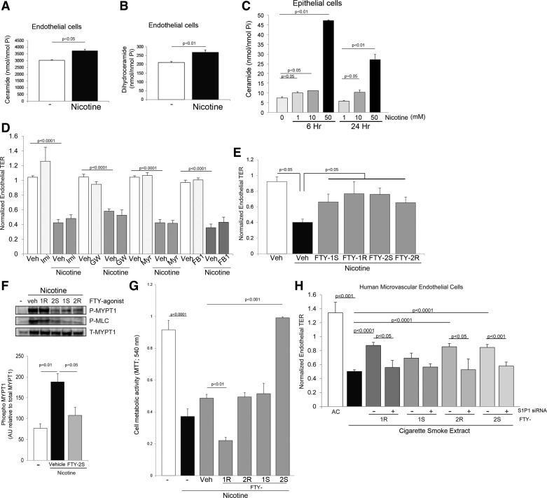
The increased use of inhaled nicotine via e-cigarettes has unknown risks to lung health. Having previously shown that cigarette smoke (CS) extract disrupts the lung microvasculature barrier function by endothelial cell activation and cytoskeletal rearrangement, we investigated the contribution of nicotine in CS or e-cigarettes (e-Cig) to lung endothelial injury. Primary lung microvascular endothelial cells were exposed to nicotine, e-Cig solution, or condensed e-Cig vapor (1-20 mM nicotine) or to nicotine-free CS extract or e-Cig solutions. Compared with nicotine-containing extract, nicotine free-CS extract (10-20%) caused significantly less endothelial permeability as measured with electric cell-substrate impedance sensing. Nicotine exposures triggered dose-dependent loss of endothelial barrier in cultured cell monolayers and rapidly increased lung inflammation and oxidative stress in mice. The endothelial barrier disruptive effects were associated with increased intracellular ceramides, p38 MAPK activation, and myosin light chain (MLC) phosphorylation, and was critically mediated by Rho-activated kinase via inhibition of MLC-phosphatase unit MYPT1. Although nicotine at sufficient concentrations to cause endothelial barrier loss did not trigger cell necrosis, it markedly inhibited cell proliferation. Augmentation of sphingosine-1-phosphate (S1P) signaling via S1P1 improved both endothelial cell proliferation and barrier function during nicotine exposures. Nicotine-independent effects of e-Cig solutions were noted, which may be attributable to acrolein, detected along with propylene glycol, glycerol, and nicotine by NMR, mass spectrometry, and gas chromatography, in both e-Cig solutions and vapor. These results suggest that soluble components of e-Cig, including nicotine, cause dose-dependent loss of lung endothelial barrier function, which is associated with oxidative stress and brisk inflammation.

Keywords: cell proliferation; inflammation; permeability; sphingosine-1-phosphate; tobacco.


Figures

Fig. 1.
Fig. 1.
Effect of nicotine on lung endothelial and epithelial barrier function. A: transcellular electrical resistance (TER) measured at the indicated time point normalized to TER at baseline (at the beginning of the measurement, before any treatment) in cells exposed to ambient air control extract (AC), nicotine-containing cigarette smoke extract (CS), or nicotine-free CS extract (all solutions were 10% vol:vol) measured by electrical cellular impedance sensing (ECIS) in primary lung microvascular endothelial cells. Values are means ± SE, n = 4–10, one-way ANOVA (with Tukey's post hoc testing for intergroup comparisons). B–D: normalized TER measured at the indicated time (hours) in primary lung rat microvascular endothelial cells (RLEC, B), primary mouse lung endothelial cells (C), and primary human lung microvascular endothelial cells (D) exposed to the indicated concentrations of nicotine. Values are means ± SE, n = 5–56, one-way ANOVA with Tukey's post hoc testing.
Fig. 2.
Fig. 2.
Effect of commercial electronic cigarette (e-Cig) solutions on lung endothelial barrier. A and B: normalized TER measured in cells (RLEC in A and human lung microvascular endothelial cells in B) exposed to nicotine (15 mM, 5 h), to CS extract (CSE with similar nicotine content), or to e-Cig extracts or condensed vapor (commercial preparation with the indicated nicotine content; 5 h). Values are means ± SE, n = 4–10, one-way ANOVA with Tukey's post hoc testing. C: normalized TER measured in human lung microvascular endothelial cells exposed to the indicated volume (microliters, μl) of e-Cig or condensed e-Cig vapor. Values are means ± SE n = 4–10, one-way ANOVA with Tukey's post hoc testing.
Fig. 3.
Fig. 3.
Composition of e-Cig and condensed e-Cig vapor. A: spectra from indicated solutions analyzed with NMR [resonances are ± 0.05 parts per million (ppm)], which detected methanol solvent OH 4.87, s; 3.30, quintet; nicotine, H2 8.50, d; H6 8.44, dd; H4 7.85, dt; H5 7.42, dd; H9a 3.24, t; H7 3.20, dd; H9b 2.37, dd; H11a 2.26, m; HN-methyl 2.17, s; H10a 1.98, m; H10b 1.88, m; H11b 1.77, m; propylene glycol H2, 3.78, m; H1 3.42, d; H3 1.15, d; glycerol, H2 3.66, tt; and H1,3 3.57, dm. In some spectra, a small aldehyde singlet (presumed acrolein) is visible at 9.77 ppm. Spectra from noted molecules were obtained from high-resolution electrospray ionization-mass spectrometry (ESI-MS, B) or gas chromatography (C) analyses of indicated solutions.
Fig. 4.
Fig. 4.
Effect of nicotine on proliferation of lung endothelial cells. Cell proliferation was determined with the metabolic activity indicator, MTT (A), or the cell division marker, CCK-8 (B), in primary rat lung microvascular endothelial cells exposed to increasing concentrations of nicotine or e-Cig (C) solutions. Values are means ± SE, n = 3, one-way ANOVA with Tukey's post hoc testing.
Fig. 5.
Fig. 5.
Oxidative stress induced by nicotine. A: nitrotyrosine levels from the plasma of C57Bl/6 mice nebulized with one dose of nicotine and harvested immediately. Levels of 8-OHdG in plasma (B) or bronchoalveolar lavage fluid (BALF, C) of C57Bl/6 mice nebulized with one dose of nicotine and collected immediately. Values are means ± SE, n = 3 per group, Student's t-test. D: detection of reactive oxygen species (ROS, green) in rat lung microvascular endothelial cells exposed to nicotine (10 mM for 30 min) with or without N-acetylcysteine (NAC, 0.5 M) using Image-iT LIVE Green Reactive Oxygen Species Detection Kit and DAPI staining of nuclei (blue).
Fig. 6.
Fig. 6.
Signaling in nicotine-induced endothelial barrier dysfunction. A: normalized TER measured in rat lung endothelial cells exposed to CS (10%) or to nicotine (15 mM) for the indicated time (hours), and effect of the antioxidant N-acetylcysteine (NAC, 0.5 M, means ± SE, n = 2–12). B: p38 MAPK activation by nicotine in lung endothelium detected by Western blot analysis for phospho- and total p38 (α, β, γ, and δ isoforms) in rat lung microvascular endothelial cells (RLEC) exposed to ambient air control extract (AC), CS extract (CS), or nicotine solution at the indicated concentrations and time points. The blot is representative of n = 3. C: normalized TER measured in RLEC exposed to nicotine (15 mM) for the indicated time (hours), and effect of a p38 inhibitor (SB203580, 30 μM, means ± SE, n = 6–50), one-way ANOVA with Tukey's post hoc test. D: myosin light chain kinase activation detected by immunoblotting for phospho-myosin light chain (Ser19) of RLEC following exposure to nicotine solution (15 mM for 1 h) in the absence or presence of the antioxidant NAC (0.5 M), the ERK-MAPK inhibitor PD98059 (PD 50 μM), or the p38 inhibitor SB203580 (SB, 30 μM). E: myosin phosphatase inhibition detected by phosphorylation of myosin phosphatase target subunit 1 (MYPT1) in RLEC exposed to nicotine (15 mM) for the indicated time points in the presence of 3 μM Rho kinase inhibitor Y29632 (RhoKinh). F: normalized TER measured in RLEC exposed to nicotine (10 mM) for the indicated time, and effect of a Rho kinase inhibitor (Y29632, 3 μM). Values are means ± SE, n = 10, one-way ANOVA with Tukey's post hoc test.
Fig. 7.
Fig. 7.
Role of sphingolipids in cellular responses to nicotine. Ceramide (A) and dihydroceramide (B) levels in RLEC following exposure to nicotine (15 mM for 4 h) and in human lung epithelial cells Beas-2B (C) following exposure to the indicated nicotine concentrations (mM) for the indicated time. Values are means ± SE, n = 3, Student's t-test. D: effect of ceramide synthesis inhibitors including that of ASMase with imipramine (Imi, 50 μM), of nSMase with GW4869 (GW, 15 μM), of the de novo pathway with myriocin (Myr, 50 nM); or of ceramide synthases in the recycling pathway with fumonisin B1 (FB1, 5 μM); or their respective vehicle controls dH2O (for FB1, Imi, and Myr) or DMSO (for GW). E: normalized TER of RLEC exposed to nicotine (15 mM for 5 h) and impact of S1P receptor agonists (FTY phosphonate analogs 1S, 1R, 2S, 2R, 10 μM) or vehicle (methanol). Values are means ± SE, n = 15–45, Student's t-test. F: myosin phosphatase inhibition MLCK activation and MLCK activation detected by phospho-Mypt1 (Thr 696) and phospho-MLC (Ser19) immunoblotting followed by densitometry in RLEC exposed to nicotine (15 mM for 20 min) and vehicle (methanol) or the indicated FTY720 analog (10 μM). Values are means ± SE, n = 3, one-way ANOVA with Tukey's post hoc test. G: cell proliferation measured with MTT in RLEC exposed to nicotine (15 mM) in the presence or absence of S1P receptor agonists (FTY phosphonate analogs 1S, 1R, 2S, 2R, 10 μM) or vehicle (methanol). Values are means ± SE, n = 3. H: TER of primary human lung microvascular cells exposed to CS (3%) or air extract (3%) and attenuated with S1P receptor agonists (FTY phosphonate analogs 1S, 1R, 2S, 2R, 10 μM) in the presence or absence of S1PR1-specific siRNA. Values are means ± SE, n = 4–14, ANOVA with Tukey's post hoc test.
Fig. 8.
Fig. 8.
Schematic of signaling events detected in lung endothelial cells exposed to nicotine. Arrows indicate activation and blocked lines indicate inhibition. Nicotine activates Rho kinase, which in turn inhibits the myosin phosphatase target subunit 1, MYPT1, enhancing phosphorylation of myosin light chains (MLC-P) to increase endothelial permeability. Rho kinase may have other targets in the cell to increase endothelial permeability because nicotine-induced oxidative stress (ROS)-dependent p38 MAPK activation also contributed to myosin light chain phosphorylation (MLC-P), but not sufficiently to alone increase permeability. Nicotine also increases the ceramide/sphingosine-1-phosphate (S1P) ratios, which may inhibit lung endothelial cell proliferation. Enhancing S1P signaling opposes the decreased cell proliferation and the increase in permeability induced by nicotine in part by inhibiting MLC phosphorylation and restoring the lung endothelial barrier function.

Similar articles

Cited by

References

    1. An N, Andrukhov O, Tang Y, Falkensammer F, Bantleon HP, Ouyang X, Rausch-Fan X. Effect of nicotine and porphyromonas gingivalis lipopolysaccharide on endothelial cells in vitro. PloS One 9: e96942, 2014. - PMC - PubMed
    1. Beckers CM, Knezevic N, Valent ET, Tauseef M, Krishnan R, Rajendran K, Corey Hardin C, Aman J, van Bezu J, Sweetnam P, van Hinsbergh VW, Mehta D, van Nieuw Amerongen GP. ROCK2 primes the endothelium for vascular hyperpermeability responses by raising baseline junctional tension. Vascul Pharmacol. First published April 11, 2015; doi:10.1016/j.vph.2015..03.017. - DOI - PMC - PubMed
    1. Brown MB, Hunt WR, Noe JE, Rush NI, Schweitzer KS, Leece TC, Moldobaeva A, Wagner EM, Dudek SM, Poirier C, Presson RG Jr, Gulbins E, Petrache I. Loss of cystic fibrosis transmembrane conductance regulator impairs lung endothelial cell barrier function and increases susceptibility to microvascular damage from cigarette smoke. Pulm Circ 4: 260–268, 2014. - PMC - PubMed
    1. Cheadle GA, Costantini TW, Bansal V, Eliceiri BP, Coimbra R. Cholinergic signaling in the gut: a novel mechanism of barrier protection through activation of enteric glia cells. Surg Infect (Larchmt) 15: 387–393, 2014. - PubMed
    1. Diab KJ, Adamowicz JJ, Kamocki K, Rush NI, Garrison J, Gu Y, Schweitzer KS, Skobeleva A, Rajashekhar G, Hubbard WC, Berdyshev EV, Petrache I. Stimulation of sphingosine 1-phosphate signaling as an alveolar cell survival strategy in emphysema. Am J Respir Crit Care Med 181: 344–352, 2010. - PMC - PubMed

Publication types

MeSH terms