. 2015 Sep;97(3):210-20.
doi: 10.1016/j.ijpsycho.2015.05.004. Epub 2015 May 12.

Spatial and temporal resolutions of EEG: Is it really black and white? A scalp current density view

Affiliations

Spatial and temporal resolutions of EEG: Is it really black and white? A scalp current density view

Borís Burle et al. Int J Psychophysiol. 2015 Sep.

Abstract

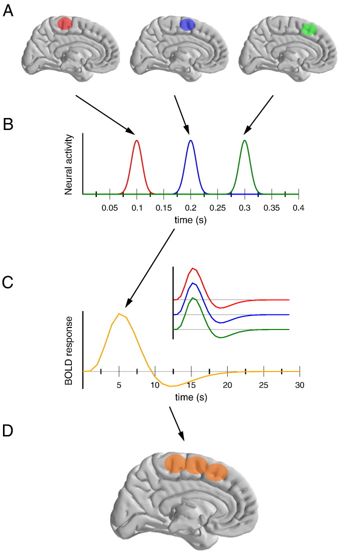
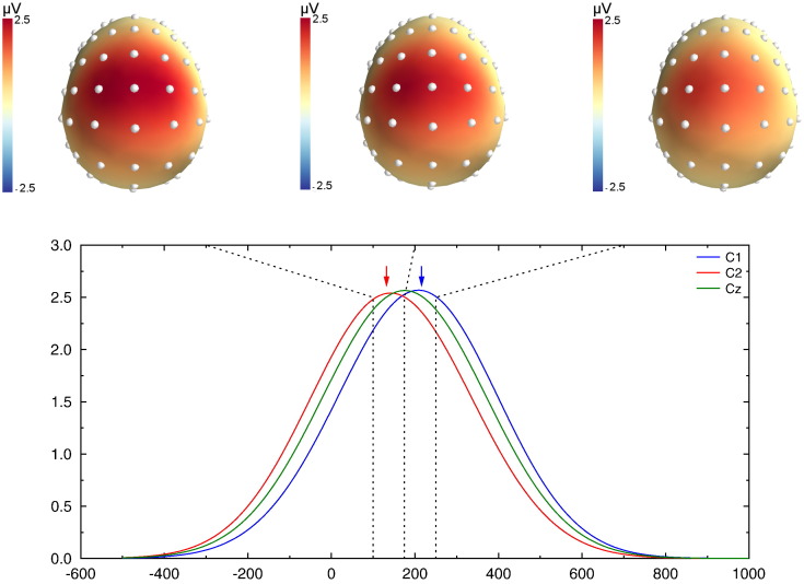
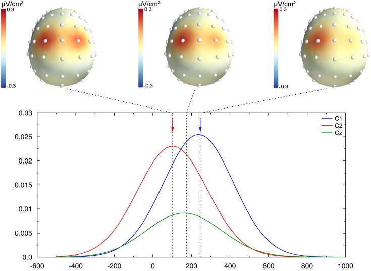
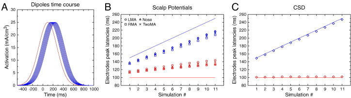
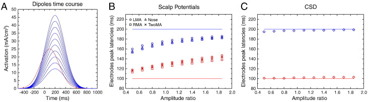
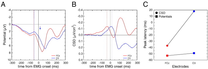
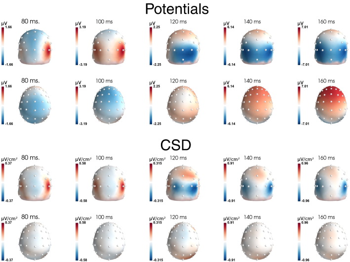
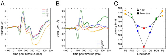
Among the different brain imaging techniques, electroencephalography (EEG) is classically considered as having an excellent temporal resolution, but a poor spatial one. Here, we argue that the actual temporal resolution of conventional (scalp potentials) EEG is overestimated, and that volume conduction, the main cause of the poor spatial resolution of EEG, also distorts the recovered time course of the underlying sources at scalp level, and hence degrades the actual temporal resolution of EEG. While Current Source Density (CSD) estimates, through the Surface Laplacian (SL) computation, are well known to dramatically reduce volume conduction effects and hence improve EEG spatial resolution, its positive impact on EEG temporal resolution is much less recognized. In two simulation studies, we first show how volume conduction and reference electrodes distort the scalp potential time course, and how SL transform provides a much better spatio-temporal description. We then exemplify similar effects on two empirical datasets. We show how the time courses of the scalp potentials mis-estimate the latencies of the relevant brain events and that CSD provides a much richer, and much more accurate, view of the spatio-temporal dynamics of brain activity.

Keywords: Current Source Density; EEG; Time resolution.


Figures

Fig. 1
Fig. 1
Schematic representation of the interaction between temporal and spatial resolutions in brain imaging studies, for example fMRI. Let's assume three neighboring regions (panel A) that are activated independently and sequentially (panel B). Their corresponding BOLD response will be identical (panel C). As a consequence, the activations of these three regions will not be separable. The resulting activation pattern (panel D) is much less refined than the actual activated areas (panel A), hence degrading the actual spatial resolution of the technique.
Fig. 2
Fig. 2
Simulation model. A–D. Graphic representation of the four meshes used as interface. A. Interface between CSF and brain. B. Between CSF and inner skull. C. Between outer skull and scalp. D. Between scalp and air. E–F. Location of the simulated dipoles, represented inside the head model. E. Top view. F. Lateral view.
Fig. 3
Fig. 3
Simulated potentials for the last simulation (100 ms vs. 250 ms). The upper row shows the scalp potential topographies obtained at 150, 175 and 250 ms. Potentials present a large central positivity. Lower panel: time course of the reconstructed potentials at electrodes C1 (blue), Cz (green) and C2 (red). The blue and red arrows indicate the latency of the recovered peak on C1 and C2. The recovered latencies on electrodes are pretty far from the underlying dipole ones. One can also note the large activity observed on Cz, despite the absence of direct underlying dipole.
Fig. 4
Fig. 4
CSD transform of the data presented in Fig. 3. The topographies (upper row) are much better resolved, with two clear peaks of activity above the underlying dipoles. As indicated by the two colored arrows, the peak latencies of the recovered CSD nicely fit the underlying dipole peak. The recovered activity over Cz is of much lower amplitude than on C1 and C2.
Fig. 5
Fig. 5
A. Time course of the simulated cortical dipoles. B. Peak latencies of the simulated dipoles (solid lines), and recovered potential peak latencies for each reference frame (LMA: left mastoid; RMA: right mastoid; TwoMA: linked mastoids). It is clear that the latency of the right (earliest, in red) dipole is overestimated, while the latency of the left (latest, in blue) one is underestimated, whatever the reference. C. Same information after CSD transform: The recovered latencies are in close agreement with the underlying dipole time courses.
Fig. 6
Fig. 6
A. Time courses of the simulated cortical dipoles. B. Peak latencies of the simulated dipoles (solid lines) for each dipole–amplitude ratio, and recovered potential peak latencies (empty symbols) for each reference electrode. Despite a constant time course of the underlying dipoles, the recovered scalp latencies differ dramatically as a function of the amplitude ratio. C. Same information after CSD transform: The recovered latencies are in close agreement with the underlying dipole time course.
Fig. 7
Fig. 7
Empirical data of dataset # 1: A: scalp potential time course recorded by electrodes C3 and FCz in a manual reaction time task (right response), time-locked to the response-EMG onset. The time courses are pretty similar between the two electrodes, and mainly present a large and temporally spread positivity that peaks slightly after EMG onset. A small bump is observed around − 50 ms, with a similar time course for both electrodes. B. Same data after SL computation: The recovered activities dramatically differ between the two electrodes. Over electrode FCz, a negative peak is observed around 40 ms before, and resolves shortly after, EMG onset. Over C3, a negative activity peaks shortly after EMG onset (around 20 ms). Panel C summarizes the obtained latencies (y-axis) as a function of the electrodes (x-axis, red: FCz, blue: C3), for scalp potentials (colored squares) and CSD (colored circles).
Fig. 8
Fig. 8
Topographies obtained on dataset # 2. The first two rows present the scalp potential topographies, viewed from back (first row) and top (second row), from 80 to 160 ms. The two lowest rows present the same data, at the same latencies, after CSD computation.
Fig. 9
Fig. 9
Time courses obtained from dataset # 2. A. Time course of the negative scalp potential at the selected occipital and parietal electrodes. Only the left electrodes are shown. Activities over the different electrodes present a very similar shape and time course. B. Same data after SL transform: the shape and the timing of the activities dramatically differ between electrodes. C. Summary of the peak latencies (y-axis) for the representative electrodes (x-axis) for scalp potentials (colored squares) and CSD (colored circles). While the latency differences are rather small for scalp potential data (max difference: 38 ms), the CSD evidences a clear occipito-parietal latency gradient (max difference: 81 ms).

References

    1. Babiloni F., Babiloni C., Fattorini L., Carducci F., Onorati P., Urbano A. Performances of surface Laplacian estimators: a study of simulated and real scalp potential distributions. Brain Topogr. 1995;8(1):35–45. - PubMed
    1. Babiloni F., Cincotti F., Carducci F., Rossini P.M., Babiloni C. Spatial enhancement of EEG data by surface Laplacian estimation: the use of magnetic resonance imaging-based head models. Clin. Neurophysiol. 2001;112(5):724–727. (May) - PubMed
    1. Bonini F., Burle B., Liégeois-Chauvel C., Régis J., Chauvel P., Vidal F. Action monitoring and medial frontal cortex: leading role of supplementary motor area. Science. 2014;343(6173):888–891. (Feb) - PubMed
    1. Burle B., Roger C., Allain S., Vidal F., Hasbroucq T. Error negativity does not reflect conflict: a reappraisal of conflict monitoring and anterior cingulate cortex activity. J. Cogn. Neurosci. 2008;20(9):1637–1655. (Sep) - PubMed
    1. Burle B., Vidal F., Tandonnet C., Hasbroucq T. Physiological evidence for response inhibition in choice reaction time tasks. Brain Cogn. 2004;56(2):153–164. (Nov) - PubMed

Publication types

LinkOut - more resources