

CSI 3.0: a web server for identifying secondary and super-secondary structure in proteins using NMR chemical shifts
- PMID: 25979265
- PMCID: PMC4489240
- DOI: 10.1093/nar/gkv494
CSI 3.0: a web server for identifying secondary and super-secondary structure in proteins using NMR chemical shifts
Abstract
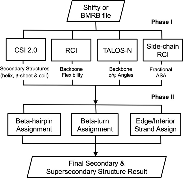
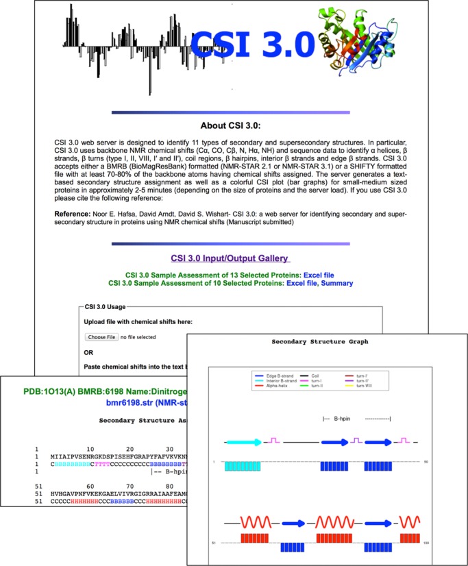
The Chemical Shift Index or CSI 3.0 (http://csi3.wishartlab.com) is a web server designed to accurately identify the location of secondary and super-secondary structures in protein chains using only nuclear magnetic resonance (NMR) backbone chemical shifts and their corresponding protein sequence data. Unlike earlier versions of CSI, which only identified three types of secondary structure (helix, β-strand and coil), CSI 3.0 now identifies total of 11 types of secondary and super-secondary structures, including helices, β-strands, coil regions, five common β-turns (type I, II, I', II' and VIII), β hairpins as well as interior and edge β-strands. CSI 3.0 accepts experimental NMR chemical shift data in multiple formats (NMR Star 2.1, NMR Star 3.1 and SHIFTY) and generates colorful CSI plots (bar graphs) and secondary/super-secondary structure assignments. The output can be readily used as constraints for structure determination and refinement or the images may be used for presentations and publications. CSI 3.0 uses a pipeline of several well-tested, previously published programs to identify the secondary and super-secondary structures in protein chains. Comparisons with secondary and super-secondary structure assignments made via standard coordinate analysis programs such as DSSP, STRIDE and VADAR on high-resolution protein structures solved by X-ray and NMR show >90% agreement between those made with CSI 3.0.
© The Author(s) 2015. Published by Oxford University Press on behalf of Nucleic Acids Research.
Figures
References
-
- Richardson J.S. The anatomy and taxonomy of protein structure. Adv. Protein Chem. 1981;34:167–339. - PubMed
-
- Wuthrich K. NMR of Proteins and Nucleic Acids. NY: John Wiley and Sons; 1986.
-
- Wuthrich K. Protein structure determination in solution by NMR spectroscopy. J. Biol. Chem. 1990;265:22059–22062. - PubMed
-
- Wishart D.S., Sykes B.D., Richards F.M. The chemical shift index: a fast and simple method for the assignment of protein secondary structure through NMR spectroscopy. Biochemistry. 1992;31:1647–1651. - PubMed
-
- Wishart D.S., Sykes B.D. The 13C chemical shift index: a simple method for the identification of protein secondary structure using 13C chemical shift data. J. Biomol. NMR. 1994;4:171–180. - PubMed
Publication types
MeSH terms
LinkOut - more resources
Full Text Sources
Other Literature Sources
