. 2015 Jul 10:209:317-26.
doi: 10.1016/j.jconrel.2015.05.267. Epub 2015 May 13.

Cancer cell surface induced peptide folding allows intracellular translocation of drug

Affiliations

Cancer cell surface induced peptide folding allows intracellular translocation of drug

Scott H Medina et al. J Control Release. .

Abstract

Many lead molecules identified in drug discovery campaigns are eliminated from consideration due to poor solubility and low cell permeability. These orphaned molecules could have clinical value if solubilized and delivered properly. SVS-1 is a de novo designed peptide that preferentially folds at the surface of tumor cells, adopting a β-hairpin conformation that rapidly translocates into the cytoplasm, and ultimately nucleus, of cells. SVS-1 is stable in serum and small molecules attached to the peptide are effectively delivered to cancer cells via mechanisms involving physical translocation and, to a lesser extent, clathrin-dependent endocytosis. For example, ligating the model hydrophobic drug Paclitaxel (PTX) to SVS-1 improved its aqueous solubility by ~1000-fold and successfully delivered and released PTX to cancer cells in vitro and tumors in vivo without toxic adjuvants. These results suggest that SVS-1 can serve as a simple, effective delivery platform for molecules with poor solubility and permeability.

Keywords: Biodistribution; Cell-penetrating peptide; Drug delivery; In vivo efficacy; Paclitaxel; Translocation.


Figures

Fig. 1
Fig. 1
Design of the SVS-1-Paclitaxel conjugate. SVS-1 peptide (green) is conjugated to paclitaxel (PTX) (red) through a PEG spacer (blue) and self-immolative di-sulfide linker (purple). The PTX-S-S-PEG-SVS-1 conjugate electrostatically engages the negatively charged surface components of cancer cells, promoting folding of the peptide into a bioactive β-hairpin. This folding event allows the peptide carrier to interpolate into the lipid bilayer leading to translocation of the conjugate into the cancer cell cytoplasm. Intracellular glutathione then reduces the self-immolative disulfide linkage leading to release of free PTX and cancer cell death.
Fig. 2
Fig. 2
(Top) Live-cell imaging of A549 cells after addition of 5 µM fluorescein-labeled SVS-1 peptide, as a function of time (scale bar = 10 µm). Associated z-stacks are also shown. (Bottom) Percentage of total cellular fluorescence measured at the membrane, within the cytoplasm or localized at the nucleus.
Fig. 3
Fig. 3
Internalization of FI-PEG-GG-SVS-1 into (A) A549, (B) HeLa or (C) MCF-7 cancer cells, as well as (D) non-cancerous HUVECs after 1 hour incubation with SVS-1 alone (open bars), or with ATP depletion (gray bars) and hyperosmolar sucrose (hatched bars) pre-conditioning, as well as the control peptide FI-PEG-GG-SVS-2 (black bars). Uptake results under ATP depletion and hyperosmolar sucrose conditions were compared to direct SVS-1 incubation (open bars) to determine statistical significance, which is denoted by ** indicating p ≤ 0.01.
Fig. 4
Fig. 4
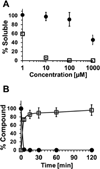
(A) Enhancement of Paclitaxel (PTX) solubility upon conjugation to the SVS-1 peptide (PTX-S-S-PEG-GG-SVS-1; ) compared to free PTX () after a 24 hour incubation in 25mM HEPES buffer (pH 7.4, 37°C). (B) Release of free PTX () and corresponding disappearance of PTX-S-S-PEG-GG-SVS-1 () on incubation of the conjugate with 5 mM glutathione at 37°C. Lines included to aid visualization.
Fig. 5
Fig. 5
Representative cytotoxicity profile of the PTX-S-S-PEG-GG-SVS-1 conjugate, free PTX or the SVS-1 peptide towards A549 (lung carcinoma) cancer cells after 72 hours of incubation. GraphPad Prism 5 software was used to fit cytotoxicity curves and, as shown in the table for all the cell lines investigated, calculate IC50 values employing a log(inhibitor) vs. response (three parameter) non-linear regression model.
Fig. 6
Fig. 6
Stability of the GG-SVS-1 () or GG-DSVS-1 () carriers, and the control Penetratin (), in the presence of human serum.
Fig. 7
Fig. 7
Time dependent tumor vs. skin fluorescence ratio following i.v. injection of the fluorescently-labeled Cy5-PEG-GG-SVS-1 peptide, it’s enantiomer Cy5-PEG-GG-DSVS-1, or the control compound Cy5-PEG-GG-NH2, to A549 tumor bearing mice.
Fig. 8
Fig. 8
(A) Percent tumor volume change for mice receiving the PTX-S-S-PEG-GG-DSVS-1 peptide-drug conjugate at the MTD (20 mg/kg), free drug (PTX) at its maximum soluble dose (1 mg/kg), or vehicle (5% EtOH, 95% HBSS). Red arrows represent the dosing regimen. Results shown as the mean ± standard error of the mean; * = p < 0.001 compared to the vehicle control. (B) Representative images of tumor tissue from mice receiving the PTX-S-S-PEG-GG-DSVS-1 peptide-drug conjugate (top), free PTX (middle), or vehicle control (bottom) (scale bar = 0.5 cm). (C) Change in percent body weight of tumor-bearing mice during treatment.

Similar articles

Cited by

References

    1. Sinthuvanich C, Veiga AS, Gupta K, Gaspar D, Blumenthal R, Schneider JP. Anticancer β-Hairpin Peptides: Membrane-Induced Folding Triggers Activity. J. Am. Chem. Soc. 2012;134:6210–6217. - PMC - PubMed
    1. Gaspar D, Veiga AS, Sinthuvanich C, Schneider JP, Castanho MARB. Anticancer Peptide SVS-1: Efficacy Precedes Membrane Neutralization. Biochemistry. 2012;51:6263–6265. - PMC - PubMed
    1. Geisow MJ. Fluorescein conjugates as indicators of subcellular pH: A critical evaluation. Exp. Cell Res. 1984;150:29–35. - PubMed
    1. Drin G, Cottin S, Blanc E, Rees AR, Temsamani J. Studies on the Internalization Mechanism of Cationic Cell-penetrating Peptides. J. Biol. Chem. 2003;278:31192–31201. - PubMed
    1. Massodi I, Bidwell Iii GL, Raucher D. Evaluation of cell penetrating peptides fused to elastin-like polypeptide for drug delivery. J. Controlled Release. 2005;108:396–408. - PubMed

Publication types

MeSH terms