. 2015 Jun 26;290(26):16031-42.
doi: 10.1074/jbc.M115.645572. Epub 2015 May 15.

Porphyromonas gingivalis-derived RgpA-Kgp Complex Activates the Macrophage Urokinase Plasminogen Activator System: IMPLICATIONS FOR PERIODONTITIS

Affiliations

Porphyromonas gingivalis-derived RgpA-Kgp Complex Activates the Macrophage Urokinase Plasminogen Activator System: IMPLICATIONS FOR PERIODONTITIS

Andrew J Fleetwood et al. J Biol Chem. .

Abstract

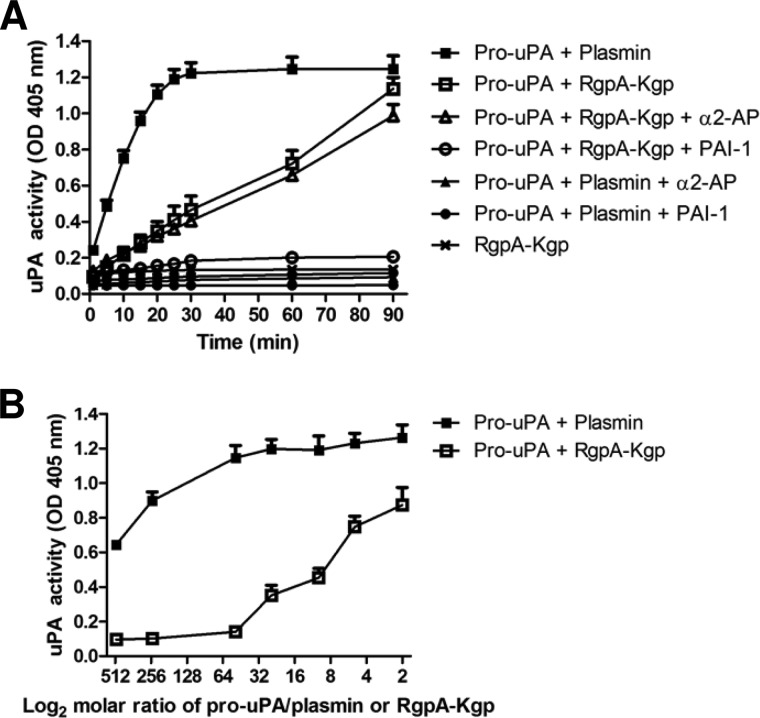
Urokinase plasminogen activator (uPA) converts plasminogen to plasmin, resulting in a proteolytic cascade that has been implicated in tissue destruction during inflammation. Periodontitis is a highly prevalent chronic inflammatory disease characterized by destruction of the tissue and bone that support the teeth. We demonstrate that stimulation of macrophages with the arginine- and lysine-specific cysteine protease complex (RgpA-Kgp complex), produced by the keystone pathogen Porphyromonas gingivalis, dramatically increased their ability to degrade matrix in a uPA-dependent manner. We show that the RgpA-Kgp complex cleaves the inactive zymogens, pro-uPA (at consensus sites Lys(158)-Ile(159) and Lys(135)-Lys(136)) and plasminogen, yielding active uPA and plasmin, respectively. These findings are consistent with activation of the uPA proteolytic cascade by P. gingivalis being required for the pathogen to induce alveolar bone loss in a model of periodontitis and reveal a new host-pathogen interaction in which P. gingivalis activates a critical host proteolytic pathway to promote tissue destruction and pathogen virulence.

Keywords: extracellular matrix; macrophage; periodontal disease; plasmin; plasminogen.


Figures

FIGURE 1.
FIGURE 1.
uPA−/− mice are protected from P. gingivalis-induced bone loss, and the RgpA-Kgp complex potentiates murine macrophage matrix degradation in a uPA-dependent manner. A, wild-type and uPA−/− mice were orally inoculated with P. gingivalis or buffer alone (uninoculated group). Alveolar bone resorption was determined, and the data are expressed as the mean ± S.D. in mm2. The data were analyzed using a one-way analysis of variance and Dunnett's T3 post hoc test (n = 12). ★, p ≤ 0.001; #, p ≤ 0.001. B, BMM (3 × 10⁴ cells/well) were placed on FITC-gelatin-coated coverslips (48 h), either untreated or in the presence of plasminogen (Plg) (500 nm), the RgpA-Kgp complex (5 nm), anti-mouse uPA mAb mU1 (267 nm), and control mAb as indicated. Cells were stained with TRITC-phalloidin (top panel, red) and DAPI (top panel, blue), and FITC-gelatin degradation (bottom panel, merged) was visualized in areas devoid of FITC staining by fluorescent microscopy at ×40 magnification. Shown are data from a representative of four independent experiments. C, quantification of FITC-gelatin degradation by BMM. The area of gelatin degradation per total cell area was determined and expressed as a percentage ± S.D. relative to untreated cells (n = 4). ★p ≤ 0.05 (Student's t test) (8). D, BMM were stimulated with the RgpA-Kgp complex (20 nm for 24 h), and the relative gene expression of murine uPA and uPAR was measured by qPCR. The data were normalized to UBC reference gene and expressed relative to untreated cells ± S.D. (n = 5).
FIGURE 2.
FIGURE 2.
Dose-dependent increase in mouse macrophage matrix degradation by RgpA-Kgp complex. A, BMM (3 × 10⁴ cells/well) were placed on FITC-gelatin-coated coverslips (48 h), in the presence of plasminogen (Plg) (500 nm) and increasing concentrations of RgpA-Kgp complex (5, 20, or 50 nm). Cells were stained with TRITC-phalloidin (top panel, red) and DAPI (top panel, blue), and FITC-gelatin degradation (bottom panel, merged) was visualized in areas devoid of FITC staining by fluorescent microscopy at ×20 magnification. Shown are data from a representative of three independent experiments. B, quantification of FITC-gelatin degradation by BMM. The area of gelatin degradation per total cell area was determined and expressed as a percentage ± S.D. relative to cells treated with Plg + RgpA-Kgp (5 nm). ★, p ≤ 0.05 (Student's t test, n = 3).
FIGURE 3.
FIGURE 3.
The RgpA-Kgp complex potentiates human macrophage matrix degradation in a uPA-dependent manner. A, MDM (3 × 10⁴ cells/well) were placed on FITC-gelatin-coated coverslips (24 h), either untreated or in the presence of plasminogen (Plg) (500 nm), RgpA-Kgp complex (5 nm), anti-human uPA mAb U-16 (267 nm), and control mAb as indicated. Cells were stained with TRITC-Phalloidin (top panel, red), DAPI (top panel, blue), and FITC-gelatin degradation (bottom panel, merged) was visualized in areas devoid of FITC staining by fluorescent microscopy at ×40 magnification. Shown are data from a representative of four independent experiments. B, quantification of FITC-gelatin degradation by MDM. The area of gelatin degradation per total cell area was determined and expressed as a percentage ± S.D. relative to untreated cells (n = 4). ★, p ≤ 0.05 (Student's t test). C, MDM were stimulated with RgpA-Kgp complex (20 nm for 24 h), and the relative gene expression of human uPA and uPAR was measured by qPCR. The data were normalized to that of the UBC reference gene and expressed relative to untreated cells ± S.D. (n = 5).
FIGURE 4.
FIGURE 4.
The RgpA-Kgp complex cleaves pro-uPA at the major consensus sites. A, pro-uPA (4.5 μm) was incubated with plasmin (50 nm) or the RgpA-Kgp complex (50 nm) for 10 min, reaction mixtures were separated by SDS-PAGE (12%) under reducing conditions, and protein bands were stained with Coomassie Brilliant Blue. Samples were labeled prior to separation by SDS-PAGE as before (see “Experimental Procedures”) (37). Indicated are intact pro-uPA (labeled P, 55 kDa) and the major fragments produced: band I (33 kDa), band II (30 kDa), band III (20 kDa), band IV (18 kDa), and band V (15 kDa). B, time-dependent cleavage of pro-uPA (250 nm) by plasmin (10 nm) or the RgpA-Kgp complex (10 nm) measured at 1, 10, 30, and 60 min by Western blot. Protein bands were detected with anti-uPA mAb specific for the protease domain of uPA. Indicated is the intact single-chain pro-uPA (labeled P, 55 kDa). Western blots were performed under reducing conditions. C, peptides from band I included amino acids from Lys136 to Leu411 of the primary human uPA sequence conforming to the sequence of LMW-uPA. The N-terminal peptide (136KPS … ELK145) was doubly acetylated, confirming that the RgpA-Kgp complex cleaved at the consensus Lys135-Lys136 site (indicated with arrowhead). D, MS/MS spectra of the most N-terminal peptide detected for band I. Although the Mascot score was low for this peptide (because of the prolines in the sequence; see main text), the fragmentation pattern was consistent with the sequence and indicated the presence of two acetyl groups at the N-terminal lysine residue. E, peptides from band II included amino acids from Ile159 to Leu411, conforming to the sequence of the uPA B-chain. The N-terminal peptide (159IIG … IYR178) was acetylated, confirming that the RgpA-Kgp complex cleaved at the consensus Lys158-Ile159 site (indicated with arrowhead). F, MS/MS spectra of the most N-terminal peptide detected for band II. This rich spectrum matched to the sequence Ac-IIGGEFTTIENQPWFAAIYR with a high Mascot score of 94. G, schematic showing the domain structure of human pro-uPA and the major cleavage sites of plasmin and RgpA-Kgp complex (indicated with arrowheads at Lys135-Lys136 and Lys158-Ile159). The A- and B-chains of uPA are held together by a single interchain disulfide bond (represented by S-S).
FIGURE 5.
FIGURE 5.
The RgpA-Kgp complex activates pro-uPA to form uPA. A, human pro-uPA activation by plasmin and RgpA-Kgp complex were compared in a kinetic chromogenic assay. Pro-uPA (50 nm) was incubated with plasmin (10 nm) or the RgpA-Kgp complex (10 nm) in the presence of the inhibitors α2-antiplasmin (α2-AP, 100 nm) or PAI-1 (100 nm). The uPA-specific substrate (0.6 mm; S2444) and absorbance was monitored for 90 min at 37 °C at 405 nm. B, comparison of the dose-dependent activation of pro-uPA (50 nm) by plasmin and the RgpA-Kgp complex (25 to 0.1 nm). The molar ratios measured were 500, 250, 50, 25, 10, 5, and 2 of pro-uPA to 1 of plasmin or RgpA-Kgp. The data are plotted on a log2 scale. Absorbance at 405 nm was measured after 60 min at 37 °C (n = 5, ± S.D.).
FIGURE 6.
FIGURE 6.
The RgpA-Kgp complex activates plasminogen to form plasmin. A, plasminogen activation by uPA and RgpA-Kgp complex were compared in a kinetic chromogenic assay. Plasminogen (250 nm) was incubated with uPA (50 nm) or RgpA-Kgp complex (50 nm) in the presence of the plasmin-specific substrate (0.5 mm; S2251), and absorbance was monitored at 405 nm for 180 min at 37 °C. B, comparison of the dose-dependent activation of plasminogen (250 nm) by uPA and the RgpA-Kgp complex (125 nm to 0.5 nm). The molar ratios measured were 500, 250, 50, 25, 10, 5, and 2 of plasminogen to 1 of uPA or RgpA-Kgp. The data are plotted on a log2 scale. Absorbance at 405 nm was measured after 120 min at 37 °C (n = 5, ± S.D.). C, uPA−/− BMM (3 × 10⁴ cells/well) were placed on FITC-gelatin-coated coverslips (48 h), in the presence of Plg (500 nm) or in the presence of Plg + RgpA-Kgp complex (5 nm). Cells were stained with TRITC-Phalloidin (top panel, red) or DAPI (top panel, blue), and FITC-gelatin degradation (bottom panel, merged) was visualized in areas devoid of FITC staining by fluorescent microscopy at ×40 magnification. Shown are data from a representative of three independent experiments. D, the area of gelatin degradation per total cell area was determined and expressed as a percentage ± S.D. relative to cells treated with plasminogen alone (n = 3). ★, p ≤ 0.05 (Student's t test).
FIGURE 7.
FIGURE 7.
Model of P. gingivalis activation of the destructive uPA/plasminogen proteolytic cascade. P. gingivalis-derived RgpA-Kgp complex activation of the uPA/plasminogen pathway (via cleavage and activation of the zymogens, pro-uPA and plasminogen, respectively) initiates a downstream proteolytic cascade promoting periodontal tissue breakdown, fibrinolysis and bleeding, complement activation, and alveolar bone loss (see “Discussion”). This provides the pathogen with a source of nutrients in the form of tissue breakdown products and hemin, contributing to its colonization and virulence, as well as promoting PD. In cells that express uPAR and plasminogen receptors (e.g. macrophages), RgpA-Kgp complex activation of the uPA/plasminogen pathway occurs at the cell surface to promote pericellular proteolysis of the extracellular matrix. RgpA-Kgp-induced activation of uPA at the cell surface may also promote uPA-uPAR signaling, which is known to play a role in cell adhesion, survival, and motility (4).

Similar articles

Cited by

References

    1. Carmeliet P., Moons L., Lijnen R., Baes M., Lemaître V., Tipping P., Drew A., Eeckhout Y., Shapiro S., Lupu F., Collen D. (1997) Urokinase-generated plasmin activates matrix metalloproteinases during aneurysm formation. Nat. Genet. 17, 439–444 - PubMed
    1. Andreasen P. A., Egelund R., Petersen H. H. (2000) The plasminogen activation system in tumor growth, invasion, and metastasis. Cell Mol. Life Sci. 57, 25–40 - PMC - PubMed
    1. Danø K., Behrendt N., Høyer-Hansen G., Johnsen M., Lund L. R., Ploug M., Rømer J. (2005) Plasminogen activation and cancer. Thromb. Haemost. 93, 676–681 - PubMed
    1. Smith H. W., Marshall C. J. (2010) Regulation of cell signalling by uPAR. Nat. Rev. Mol. Cell Biol. 11, 23–36 - PubMed
    1. Hamilton J. A., Campbell I. K., Wojta J., Cheung D. (1992) Plasminogen activators and their inhibitors in arthritic disease. Ann. N.Y. Acad. Sci. 667, 87–100 - PubMed

Publication types

MeSH terms

Substances

LinkOut - more resources