

The role of epigenetics in the endothelial cell shear stress response and atherosclerosis
- PMID: 25979369
- PMCID: PMC4592147
- DOI: 10.1016/j.biocel.2015.05.001
The role of epigenetics in the endothelial cell shear stress response and atherosclerosis
Abstract
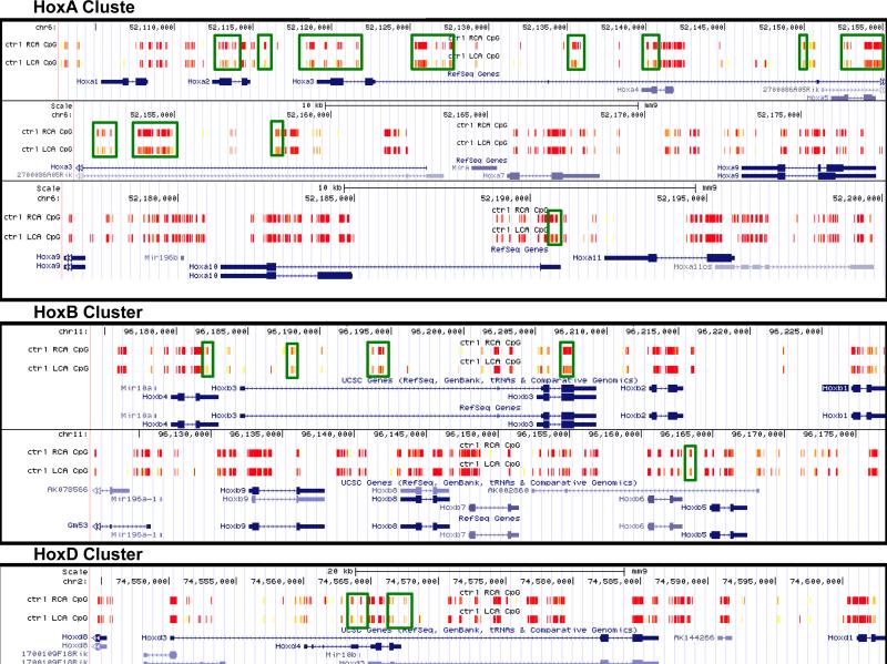
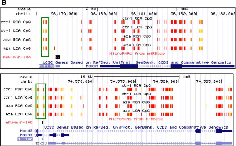
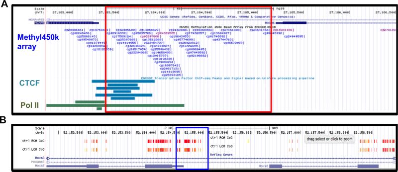
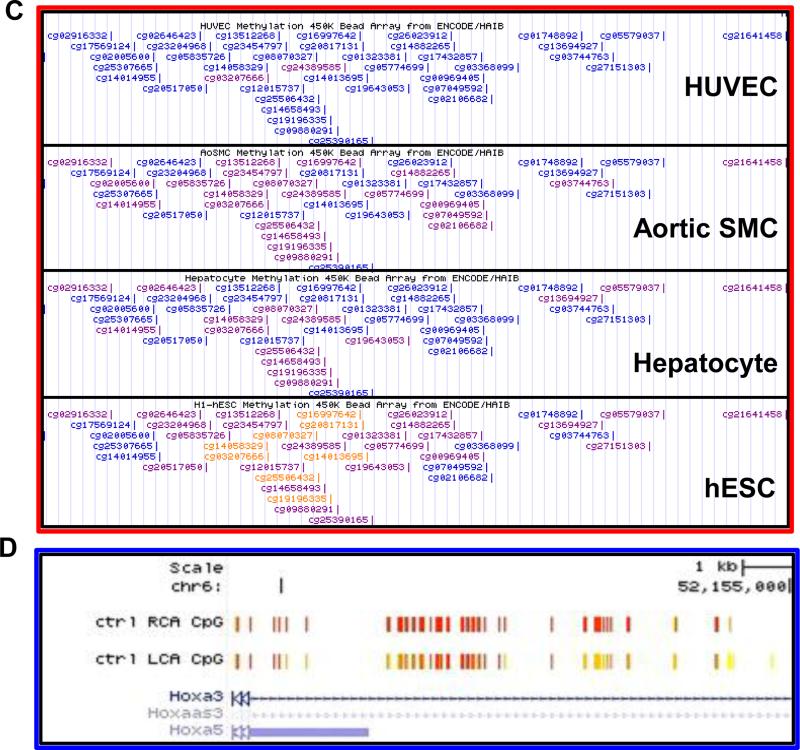
Currently in the field of vascular biology, the role of epigenetics in endothelial cell biology and vascular disease has attracted more in-depth study. Using both in vitro and in vivo models of blood flow, investigators have recently begun to reveal the underlying epigenetic regulation of endothelial gene expression. Recently, our group, along with two other independent groups, have demonstrated that blood flow controls endothelial gene expression by DNA methyltransferases (DNMT1 and 3A). Disturbed flow (d-flow), characterized by low and oscillating shear stress (OS), is pro-atherogenic and induces expression of DNMT1 both in vivo and in vitro. D-flow regulates genome-wide DNA methylation patterns in a DNMT-dependent manner. The DNMT inhibitor 5-Aza-2'deoxycytidine (5Aza) or DNMT1 siRNA reduces OS-induced endothelial inflammation. Moreover, 5Aza inhibits the development of atherosclerosis in ApoE(-/-) mice. Through a systems biological analysis of genome-wide DNA methylation patterns and gene expression data, we found 11 mechanosensitive genes which were suppressed by d-flow in vivo, experienced hypermethylation in their promoter region in response to d-flow, and were rescued by 5Aza treatment. Interestingly, among these mechanosensitive genes, the two transcription factors HoxA5 and Klf3 contain cAMP-response-elements (CRE), which may indicate that methylation of CRE sites could serve as a mechanosensitive master switch in gene expression. These findings provide new insight into the mechanism by which flow controls epigenetic DNA methylation patterns, which in turn alters endothelial gene expression, regulates vascular biology, and induces atherosclerosis. These novel findings have broad implications for understanding the biochemical mechanisms of atherogenesis and provide a basis for identifying potential therapeutic targets for atherosclerosis. This article is part of a Directed Issue entitled: Epigenetics dynamics in development and disease.
Keywords: Atherosclerosis; DNMT; Endothelial function; Epigenetic DNA methylation; Flow; Gene expression; Shear stress.
Copyright © 2015 Elsevier Ltd. All rights reserved.
Figures
Similar articles
-
Flow-dependent epigenetic DNA methylation regulates endothelial gene expression and atherosclerosis.J Clin Invest. 2014 Jul;124(7):3187-99. doi: 10.1172/JCI74792. Epub 2014 May 27. J Clin Invest. 2014. PMID: 24865430 Free PMC article.
-
Flow-Dependent Epigenetic DNA Methylation in Endothelial Gene Expression and Atherosclerosis.Arterioscler Thromb Vasc Biol. 2015 Jul;35(7):1562-9. doi: 10.1161/ATVBAHA.115.305042. Epub 2015 May 7. Arterioscler Thromb Vasc Biol. 2015. PMID: 25953647 Free PMC article. Review.
-
DNA Methyltransferase 1-Dependent DNA Hypermethylation Constrains Arteriogenesis by Augmenting Shear Stress Set Point.J Am Heart Assoc. 2017 Nov 30;6(12):e007673. doi: 10.1161/JAHA.117.007673. J Am Heart Assoc. 2017. PMID: 29191807 Free PMC article.
-
Atherosclerosis and flow: roles of epigenetic modulation in vascular endothelium.J Biomed Sci. 2019 Aug 7;26(1):56. doi: 10.1186/s12929-019-0551-8. J Biomed Sci. 2019. PMID: 31387590 Free PMC article. Review.
-
DNA methyltransferase 1 and Krüppel-like factor 4 axis regulates macrophage inflammation and atherosclerosis.J Mol Cell Cardiol. 2019 Mar;128:11-24. doi: 10.1016/j.yjmcc.2019.01.009. Epub 2019 Jan 16. J Mol Cell Cardiol. 2019. PMID: 30659837
Cited by
-
Retrospective Study of Hemodynamic Changes Before and After Carotid Stenosis Formation by Vessel Surface Repairing.Sci Rep. 2018 Apr 3;8(1):5493. doi: 10.1038/s41598-018-23842-0. Sci Rep. 2018. PMID: 29615730 Free PMC article.
-
Fluid shear stress and tumor metastasis.Am J Cancer Res. 2018 May 1;8(5):763-777. eCollection 2018. Am J Cancer Res. 2018. PMID: 29888101 Free PMC article. Review.
-
Nuclear complex of glyceraldehyde-3-phosphate dehydrogenase and DNA repair enzyme apurinic/apyrimidinic endonuclease I protect smooth muscle cells against oxidant-induced cell death.FASEB J. 2017 Jul;31(7):3179-3192. doi: 10.1096/fj.201601082R. Epub 2017 Apr 12. FASEB J. 2017. PMID: 28404743 Free PMC article.
-
Omics-based approaches to understand mechanosensitive endothelial biology and atherosclerosis.Wiley Interdiscip Rev Syst Biol Med. 2016 Sep;8(5):378-401. doi: 10.1002/wsbm.1344. Epub 2016 Jun 24. Wiley Interdiscip Rev Syst Biol Med. 2016. PMID: 27341633 Free PMC article. Review.
-
Neuroendocrinological and Epigenetic Mechanisms Subserving Autonomic Imbalance and HPA Dysfunction in the Metabolic Syndrome.Front Neurosci. 2016 Apr 14;10:142. doi: 10.3389/fnins.2016.00142. eCollection 2016. Front Neurosci. 2016. PMID: 27147943 Free PMC article. Review.
References
-
- Akimoto S, Mitsumata M, Sasaguri T, Yoshida Y. Laminar shear stress inhibits vascular endothelial cell proliferation by inducing cyclin-dependent kinase inhibitor p21(Sdi1/Cip1/Waf1). Circ Res. 2000;86:185–190. - PubMed
-
- Garcia-Cardena G, Comander JI, Blackman BR, Anderson KR, Gimbrone MA. Mechanosensitive endothelial gene expression profiles: scripts for the role of hemodynamics in atherogenesis? Ann N Y Acad Sci. 2001;947:1–6. - PubMed
-
- Chatzizisis YS, Coskun AU, Jonas M, Edelman ER, Feldman CL, Stone PH. Role of endothelial shear stress in the natural history of coronary atherosclerosis and vascular remodeling: molecular, cellular, and vascular behavior. J Am Coll Cardiol. 2007;49:2379–2393. - PubMed
-
- Chien S, Shyy JY. Effects of hemodynamic forces on gene expression and signal transduction in endothelial cells. Biol Bull. 1998;194:390–391. discussion 392-393. - PubMed
-
- He XH, Wu GF, Zhang Y, Chen XL, Zhang ZS, Zhan CY, Liu J, He JG, Xiong Y, Fang DQ, et al. [Effect of chronic enhanced external counterpulastion on gene expression profiles of arterial endothelial cells of pigs fed with high-cholesterol diet]. Nan Fang Yi Ke Da Xue Xue Bao. 2008;28:1195–1197. - PubMed
Publication types
MeSH terms
Substances
Grants and funding
LinkOut - more resources
Full Text Sources
Other Literature Sources
Medical
Miscellaneous
