. 2015 Aug;98(2):271-81.
doi: 10.1189/jlb.4A1214-577RR. Epub 2015 May 15.

Superantigens produced by catheter-associated Staphylococcus aureus elicit systemic inflammatory disease in the absence of bacteremia

Affiliations

Superantigens produced by catheter-associated Staphylococcus aureus elicit systemic inflammatory disease in the absence of bacteremia

Jin-Won Chung et al. J Leukoc Biol. 2015 Aug.

Abstract

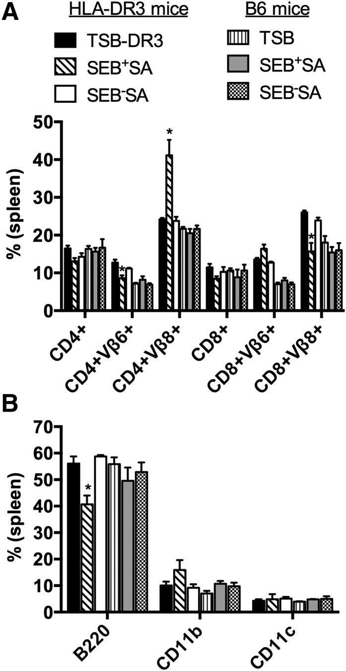
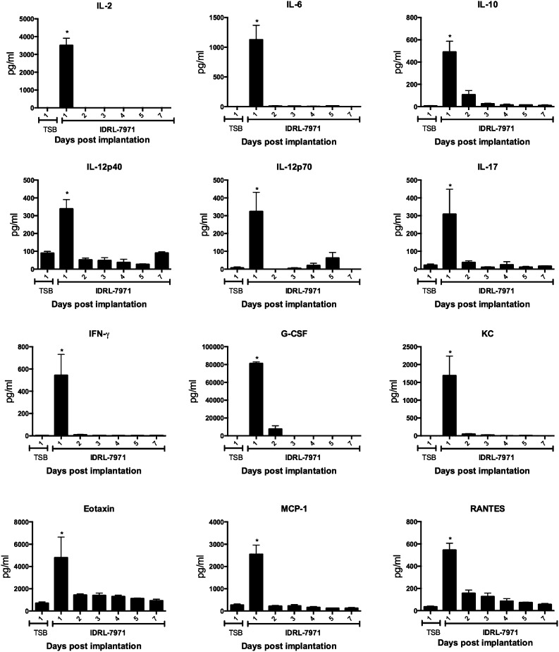
SAgs, produced by Staphylococcus aureus, play a major role in the pathogenesis of invasive staphylococcal diseases by inducing potent activation of the immune system. However, the role of SAgs, produced by S. aureus, associated with indwelling devices or tissues, are not known. Given the prevalence of device-associated infection with toxigenic S. aureus in clinical settings and the potency of SAgs, we hypothesized that continuous exposure to SAgs produced by catheter-associated S. aureus could have systemic consequences. To investigate these effects, we established a murine in vivo catheter colonization model. One centimeter long intravenous catheters were colonized with a clinical S. aureus isolate producing SAgs or isogenic S. aureus strains, capable or incapable of producing SAg. Catheters were subcutaneously implanted in age-matched HLA-DR3, B6, and AE(o) mice lacking MHC class II molecules and euthanized 7 d later. There was no evidence of systemic infection. However, in HLA-DR3 transgenic mice, which respond robustly to SSAgs, the SSAg-producing, but not the nonproducing strains, caused a transient increase in serum cytokine levels and a protracted expansion of splenic CD4(+) T cells expressing SSAg-reactive TCR Vβ8. Lungs, livers, and kidneys from these mice showed infiltration with CD4(+) and CD11b(+) cells. These findings were absent in B6 and AE(o) mice, which are known to respond poorly to SSAgs. Overall, our novel findings suggest that systemic immune activation elicited by SAgs, produced by S. aureus colonizing foreign bodies, could have clinical consequences in humans.

Keywords: HLA class II; T lymphocytes; cytokines; immunopathology; transgenic mice.


Figures

Figure 1.
Figure 1.. Efficient colonization of catheters and production of SAg by the clinical isolate S. aureus IDRL-7971.
(A) One centimeter segments of intravenous catheters were incubated with IDRL-7971, as described in Materials and Methods. The next day, catheter segments were gently rinsed in saline, longitudinally bisected, fixed in neutral buffered formalin, and processed for scanning electron microscopy. A representative image is shown. (B) Filtered culture supernatants from catheters containing IDRL-7971 were tested for the presence of SEA/B/C/D/E by the TECRA Staph Enterotoxins ID kit (3M). PC and NC, Absorbance values corresponding to positive and negative controls, respectively, provided with the kit. Representative data from 2 independent experiments are shown. (C) Splenocytes from HLA-DR3, B6, and AEo mice were cultured with TSB alone or with supernatants from IDRL-7971 catheter cultures. Splenocyte proliferation was determined by a thymidine incorporation assay. Representative data from 2 independent experiments are shown. (D) Splenocytes from HLA-DR3, B6, and AEo mice were cultured with catheters colonized with IDRL-7971 in a transwell culture plate. Supernatants were harvested, filtered, serially diluted, and tested for the presence of IL-2 by ELISA. Representative data from 2 independent experiments are shown.
Figure 2.
Figure 2.. Subcutaneous implantation of catheters colonized with SAgs-producing S. aureus IDRL-7971 causes expansion of SAg-reactive T cells.
Control catheters or catheters precolonized with IDRL-7971 were implanted subcutaneously at the backs of HLA-DR3, B6, and AEo mice. Seven days later, spleens were collected and analyzed by flow cytometry. (A) Percentage of CD4+ and CD8+ T cells expressing SEB-reactive TCR Vβ8 and the control TCR Vβ6 in the spleens. (B) Absolute numbers of CD4+ and CD8+ T cells expressing SEB-reactive TCR Vβ8 and the control TCR Vβ6 in spleens of HLA-DR3 transgenic mice implanted with control or IDRL-7971 catheters. (C) Percentages of B220+, CD11b+, and CD11c+ cells in the spleens. Each bar represents mean ± se from 4 to 8 mice. *P < 0.05 compared with corresponding TSB group.
Figure 3.
Figure 3.. Subcutaneous implantation of catheters colonized with SAg-producing S. aureus IDRL-7971 causes infiltration of vital organs with inflammatory cells.
HLA-DR3 and B6 mice were implanted subcutaneously with control catheters or catheters precolonized with IDRL-7971. Seven days later, lungs, livers, and kidneys were collected, formalin fixed, paraffin embedded, sectioned, and stained with H&E. Representative images are shown. Insets within each panel show higher magnification. Representative images are shown.
Figure 4.
Figure 4.. Subcutaneous implantation of catheters colonized with SAg-producing clinical S. aureus IDRL-7971 causes infiltration with CD4+ T cells and CD11b+ cells.
HLA-DR3 and B6 mice were implanted subcutaneously with control catheters or catheters precolonized with IDRL-7971. Livers collected 7 d later were cryosectioned and stained with FITC-conjugated CD4 (A) and CD11b (B) antibodies. Representative images are shown. (Left) DAPI stain of the nuclei; (middle) FITC stain; (right) DAPI-FITC overlay. Representative images are shown.
Figure 5.
Figure 5.. SAg toxin profile and SAg production by experimental S. aureus isolates, SEB+SA and SEBSA.
(A) Filtered culture supernatants of SEB+SA and SEBSA were tested for the presence of SEA/B/C/D/E by the TECRA Staph Enterotoxins ID kit (3M). Representative data from 2 independent experiments are shown. (B) Splenocytes from HLA-DR3, B6, and AEo mice were cultured with TSB alone or with supernatants from SEB+SA and SEBSA cultures. The extent of splenocyte proliferation was determined by thymidine incorporation assay. Representative data from several independent experiments are shown. (C) Splenocytes from HLA-DR3, B6, and AEo mice were cultured with catheters colonized with SEB+SA and SEBSA in a transwell culture plate. Supernatants were harvested, filtered, and tested for the presence of IL-2 by ELISA. Representative data from 2 independent experiments are shown.
Figure 6.
Figure 6.. Subcutaneous implantation of catheters colonized with SEB+SA and SEBSA causes expansion of SAg-reactive T cells.
Control catheters or catheters precolonized with SEB+SA and SEB-SA were implanted subcutaneously into HLA-DR3 and B6 mice. Seven days later, spleens were collected and analyzed by flow cytometry. (A) Absolute numbers of CD4+ and CD8+ T cells expressing SEB-reactive TCR Vβ8 and the control TCR Vβ6 in spleens. (B) Absolute numbers of B220+, CD11b+, and CD11c+ cells in the spleens. Each bar represents mean ± se from 4 to 8 mice. *P < 0.05 by use of the Kruskal-Wallis test, followed by Dunn’s multiple comparisons test.
Figure 7.
Figure 7.. Subcutaneous implantation of catheters colonized with S. aureus causes infiltration of vital organs with inflammatory cells in a SAg-dependent manner.
(A) B6 and (B) HLA-DR3 transgenic mice were implanted subcutaneously with control catheters or catheters precolonized with SEB+SA or SEBSA. Seven days later, lungs, livers, and kidneys were collected, formalin fixed, paraffin embedded, sectioned, and stained with H&E. Insets within each panel show higher magnification. Representative images are shown.
Figure 8.
Figure 8.. Subcutaneous implantation of catheters colonized with SEB+SA causes infiltration of vital organs with CD4+ T cells and CD11b+ cells.
HLA-DR3 were implanted subcutaneously with control catheters or catheters precolonized with SEB+SA. Kidneys and livers, collected 7 d later, were cryosectioned and stained with FITC-conjugated CD4, CD8, CD11b, and CD11c antibodies. Representative images are shown. (Top) Kidney; (Bottom) liver.
Figure 9.
Figure 9.. Temporal kinetics of cytokine/chemokine production in HLA-DR3 mice implanted with catheters colonized with S. aureus.
Age-matched HLA-DR3 mice were implanted subcutaneously with control catheters or catheters precolonized with IDRL-7971. Mice were euthanized daily for 7 d, and sera were collected immediately after euthanasia. Serum levels of indicated cytokines/chemokines were determined by multiplex assay. Each bar represents mean ± se from 4 mice. *P < 0.05 by use of Kruskal-Wallis test, followed by Dunn’s multiple comparisons test. KC, keratinocyte-derived chemokine.
Figure 10.
Figure 10.. Temporal kinetics of expansion of SAg-reactive T cells in HLA-DR3 mice implanted with catheters colonized with S. aureus.
Age-matched HLA-DR3 mice were implanted subcutaneously with control catheters or catheters precolonized with IDRL-7971. Mice were euthanized daily for 7 d, spleens were collected immediately after euthanasia, and distribution of indicated T cell subsets was determined by flow cytometry. Each bar represents mean ± se from 4 mice. POD, postoperation day. *P < 0.05 compared with corresponding TSB control group.

References

    1. O’Grady N. P., Alexander M., Burns L. A., Dellinger E. P., Garland J., Heard S. O., Lipsett P. A., Masur H., Mermel L. A., Pearson M. L., Raad I. I., Randolph A. G., Rupp M. E., Saint S.; Healthcare Infection Control Practices Advisory Committee (2011) Guidelines for the prevention of intravascular catheter-related infections. Am. J. Infect. Control 39 (4 Suppl 1) S1–S34. - PubMed
    1. Donlan R. M. (2001) Biofilms and device-associated infections. Emerg. Infect. Dis. 7, 277–281. - PMC - PubMed
    1. Hidron A. I., Edwards J. R., Patel J., Horan T. C., Sievert D. M., Pollock D. A., Fridkin S. K.; National Healthcare Safety Network Team; Participating National Healthcare Safety Network Facilities (2008) NHSN annual update: antimicrobial-resistant pathogens associated with healthcare-associated infections: annual summary of data reported to the National Healthcare Safety Network at the Centers for Disease Control and Prevention, 2006-2007. Infect. Control Hosp. Epidemiol. 29, 996–1011. - PubMed
    1. Brady R. A., Leid J. G., Calhoun J. H., Costerton J. W., Shirtliff M. E. (2008) Osteomyelitis and the role of biofilms in chronic infection. FEMS Immunol. Med. Microbiol. 52, 13–22. - PubMed
    1. Archer N. K., Mazaitis M. J., Costerton J. W., Leid J. G., Powers M. E., Shirtliff M. E. (2011) Staphylococcus aureus biofilms: properties, regulation, and roles in human disease. Virulence 2, 445–459. - PMC - PubMed

Publication types

MeSH terms