Review
. 2015 Jul;36(7):471-80.
doi: 10.1016/j.tips.2015.04.003. Epub 2015 May 13.

Inflammatory reaction after traumatic brain injury: therapeutic potential of targeting cell-cell communication by chemokines

Affiliations
Review

Inflammatory reaction after traumatic brain injury: therapeutic potential of targeting cell-cell communication by chemokines

Stefka Gyoneva et al. Trends Pharmacol Sci. 2015 Jul.

Abstract

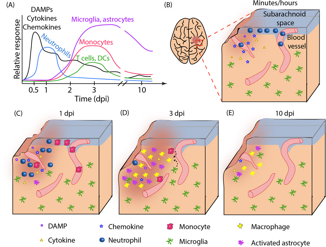
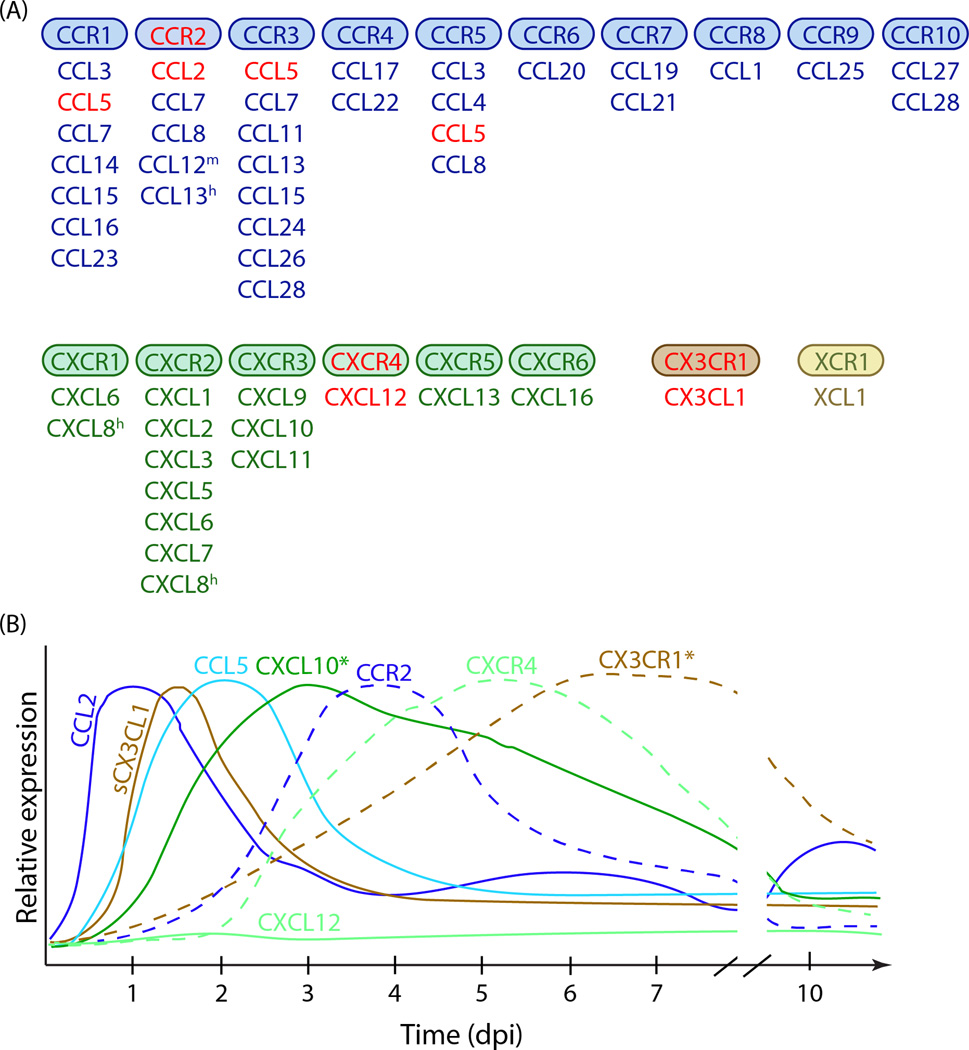
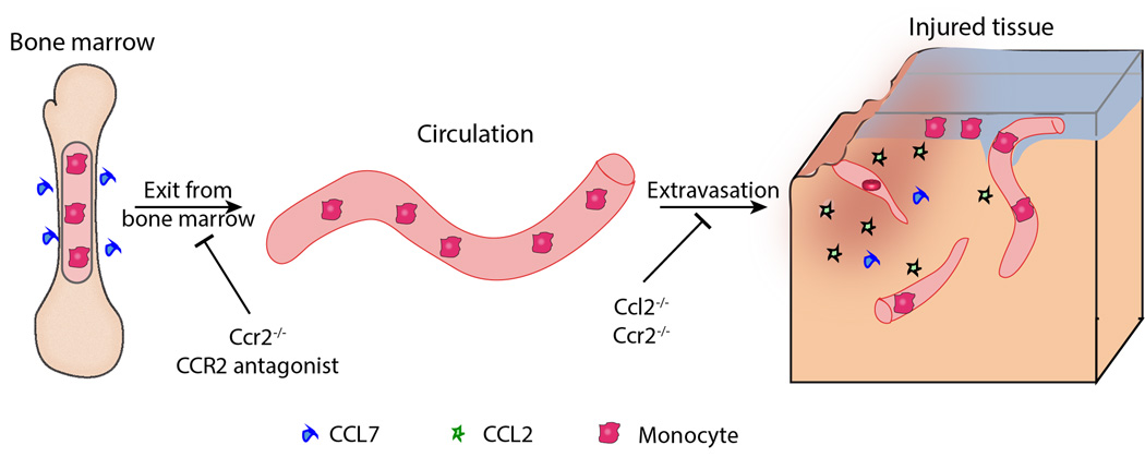
Traumatic brain injury (TBI) affects millions of people worldwide every year. The primary impact initiates the secretion of pro- and anti-inflammatory factors, subsequent recruitment of peripheral immune cells, and activation of brain-resident microglia and astrocytes. Chemokines are major mediators of peripheral blood cell recruitment to damaged tissue, including the TBI brain. Here we review the involvement of specific chemokine pathways in TBI pathology and attempts to modulate these pathways for therapeutic purposes. We focus on chemokine (C-C motif) ligand 2/chemokine (C-C motif) receptor 2 (CCL2/CCR2) and chemokine (C-X-C motif) ligand 12/chemokine (C-X-C motif) receptor 4 (CXCL12/CXCR4). Recent microarray and multiplex expression profiling have also implicated CXCL10 and CCL5 in TBI pathology. Chemokine (C-X3-C motif) ligand 1/chemokine (C-X3-C motif) receptor 1 (CX3CL1/CX3CR1) signaling in the context of TBI is also discussed. Current literature suggests that modulating chemokine signaling, especially CCL2/CCR2, may be beneficial in TBI treatment.

Keywords: CCR2; chemokines; inflammation; traumatic brain injury.


Figures

Figure 1
Figure 1. Inflammatory response to TBI
A. Time course of molecular and cellular mediators after TBI. Molecular mediators such as danger-associated molecular patterns (DAMPs), cytokines and chemokines are released immediately after injury and peak within hours. They continue to be released at later time points by the tissue damage and infiltrating cells. Neutrophil infiltration in the brain parenchyma is maximal at 1 day post injury (dpi), while monocyte, T and dendritic cell accumulation peaks at about 3 dpi. The inflammatory reaction is mostly resolved by 10 dpi. The relative response for each cells type is normalized to the maximum cellular response. B-E. Histological representation of the inflammatory reaction. B. Acute (minutes/hours) inflammation at the site of tissue lesion. A focal injury is characterized by tissue damage and secretion of DAMPs, cytokines and chemokines by the damaged cells. Neutrophils start to accumulate in the subarachnoid space (SAS) and in blood vessels (parenchymal and pial) at the injured area. C. At 1 dpi, DAMPs, cytokine and chemokine levels are reduced. Neutrophils are found throughout the parenchyma. Monocytes are being recruited to the damaged area, but most of them are still inside blood vessels. D. By 3 dpi, neutrophils are almost undetectable and monocytes predominate at the site of lesion. Astrocytes and resident microglia also assume an activated morphology. Amoeboid microglia are visually indistinguishable from monocyte-derived macrophages. Some T lymphocytes and dendritic cells are also present in the parenchyma. E. By 10 dpi, the inflammatory reaction is mostly resolved. Some activated astrocytes and macrophage-like cells can still be detected, especially in deeper brain regions (not shown).
Figure 2
Figure 2. Chemokines in the context of TBI
A. Chemokine receptor/chemokine signaling. There are four major chemokine families (CCL, CXCL, CX3CL and XCL) that bind to four families of chemokine receptors (CCR, CXCR, CX3CR and XCR). The figure shows the chemokine receptors and the ligands that have been shown to activate them. Only typical chemokine receptors that activate G proteins upon ligand binding are included. m, present only in mice; h, present only in humans. Chemokines in red are referenced in this review. B. General time course of chemokine and chemokine receptor expression in TBI. Only chemokines and receptors discussed in this review are included. The graphs show protein expression of each analyte, except for CXCL10* and CX3CR1* (*, mRNA expression shown). The relative expression for each chemokine/receptor is normalized to its own peak expression. Chemokine expression generally precedes the expression of the receptor it signals through.
Figure 3
Figure 3. CCR2 signaling in the turnover of CD11b+CD45hi monocytes
Monocytes produced in the bone marrow require CCR2 signaling – mostly activated by CCL7 – to exit the bone marrow and enter the circulation. In the presence of tissue injury, including brain injury, CD11b+CD45hiCCR2+ monocytes are recruited to the damaged area. Several chemokines, such as CCL2, CCL7, CCL8, CCL12 (mouse) and CCL13 (human) can mediate monocyte extravasation. CCR2 deletion and antagonism inhibits both monocyte egress out of the bone marrow and into tissues, while CCL2 deletion affects primarily monocyte recruitment to the site of damage.

References

    1. Faul M, et al. Traumatic Brain Injury in the United States: Emergency Department Visits, Hospitalizations and Deaths 2002–2006. Centers for Disease Control and Prevention. National Centers for Injury Prevention and Control. 2010
    1. Teasdale G, Jennett B. Assessment of coma and Impaired consciousness: A practical scale. Lancet. 1974;304(7872):81–84. - PubMed
    1. Whitnall L, et al. Disability in young people and adults after head injury: 5–7 year followup of a prospective cohort study. J Neurol Neurosurg Psychiatry. 2006;77(640–645):640. - PMC - PubMed
    1. Kiraly MA, Kiraly SJ. Traumatic Brain Injury and Delayed Sequelae: A Review -Traumatic Brain Injury and Mild Traumatic Brain Injury (Concussion) are Precursors to Later-Onset Brain Disorders, Including Early-Onset Dementia. The Scientific World Journal. 2007;7:1768–1776. - PMC - PubMed
    1. Draper K, Ponsford J. Cognitive Functioning Ten Years Following Traumatic Brain Injury and Rehabilitation. Neurophyschology. 2008;22(5):618–625. - PubMed