. 2015 Jul 15;75(14):2800-2810.
doi: 10.1158/0008-5472.CAN-14-3041. Epub 2015 May 15.

Tumor-Promoting Desmoplasia Is Disrupted by Depleting FAP-Expressing Stromal Cells

Affiliations

Tumor-Promoting Desmoplasia Is Disrupted by Depleting FAP-Expressing Stromal Cells

Albert Lo et al. Cancer Res. .

Abstract

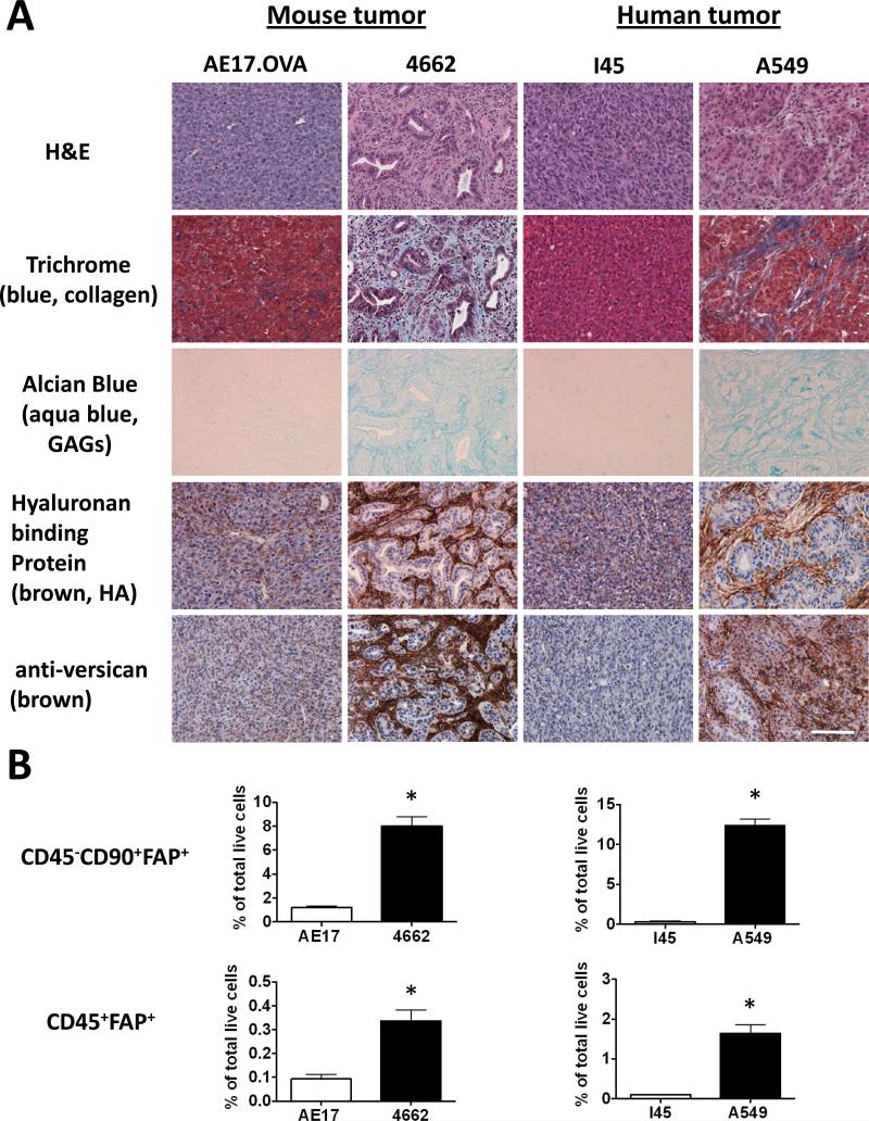
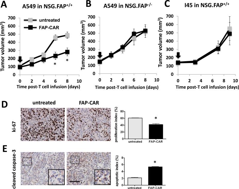
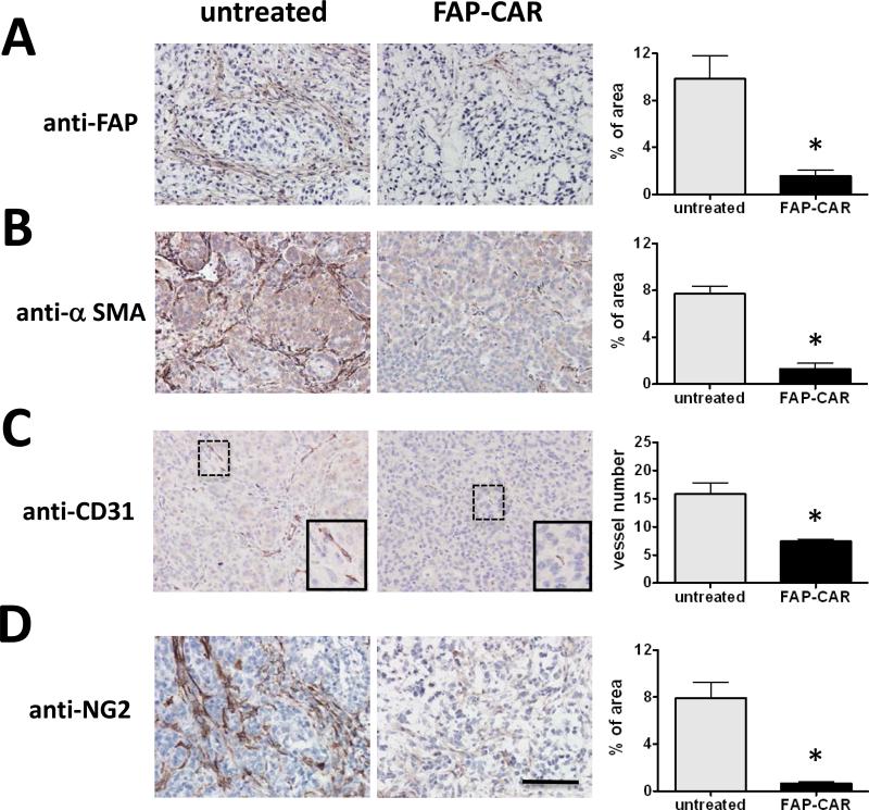
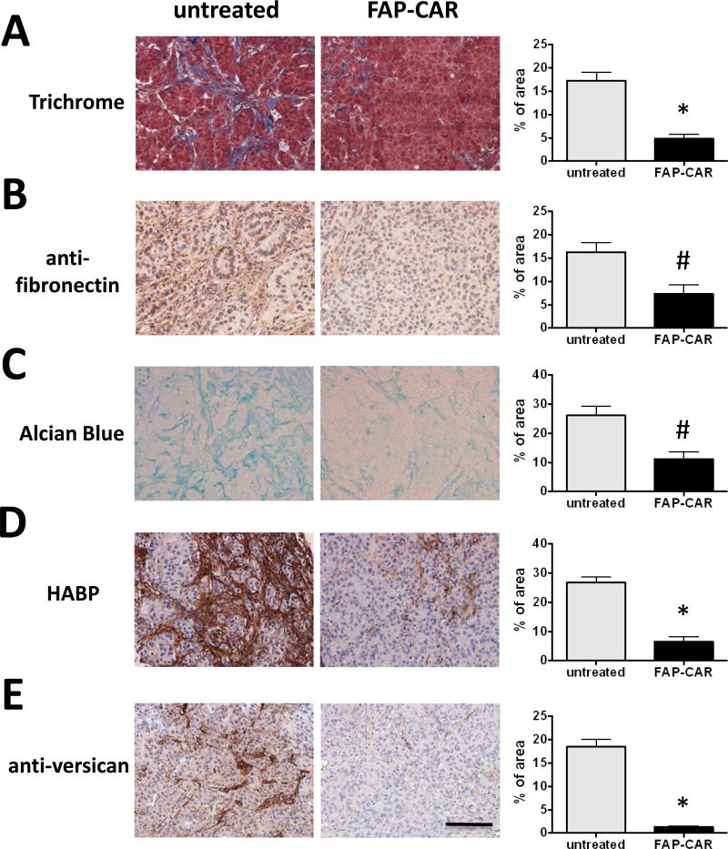
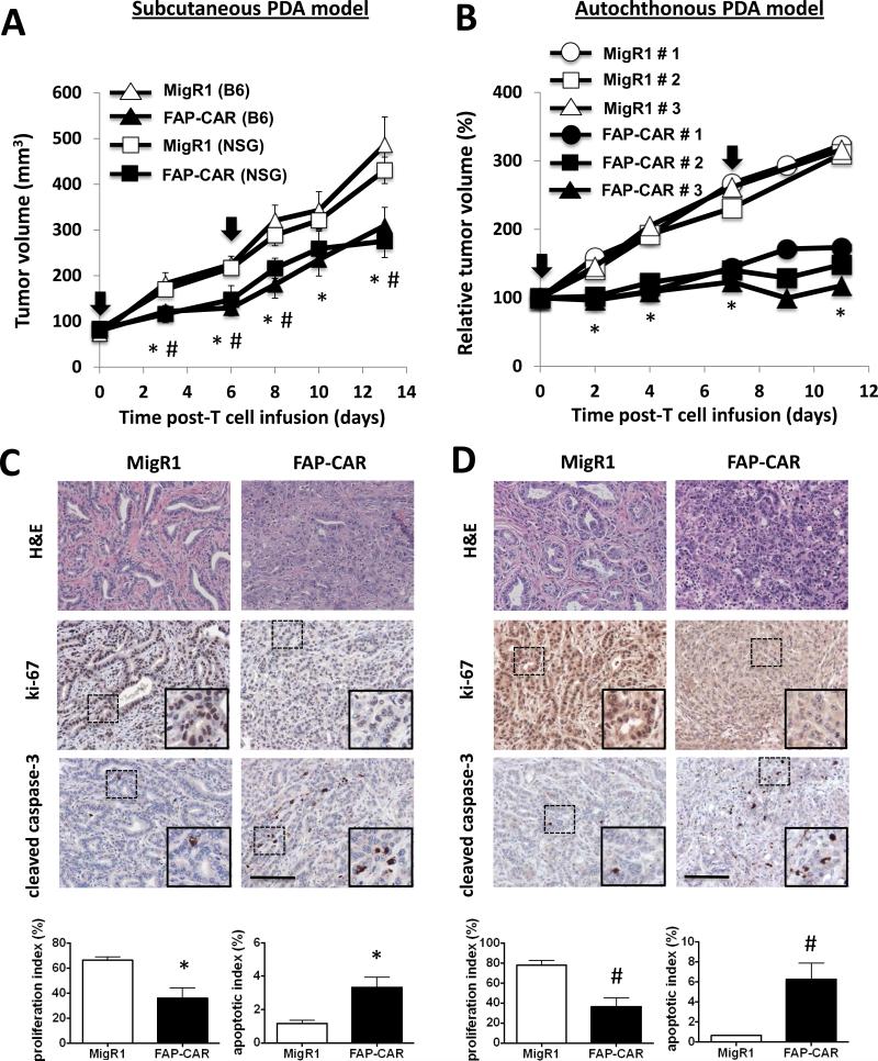
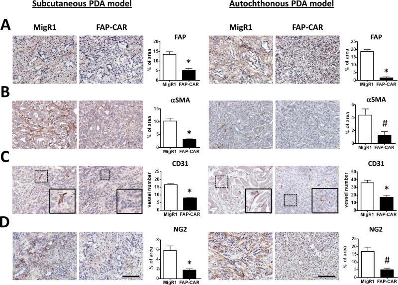
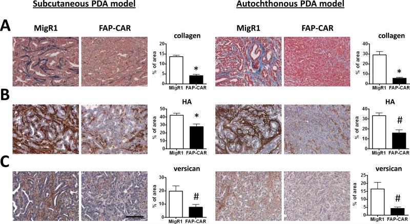
Malignant cells drive the generation of a desmoplastic and immunosuppressive tumor microenvironment. Cancer-associated stromal cells (CASC) are a heterogeneous population that provides both negative and positive signals for tumor cell growth and metastasis. Fibroblast activation protein (FAP) is a marker of a major subset of CASCs in virtually all carcinomas. Clinically, FAP expression serves as an independent negative prognostic factor for multiple types of human malignancies. Prior studies established that depletion of FAP(+) cells inhibits tumor growth by augmenting antitumor immunity. However, the potential for immune-independent effects on tumor growth have not been defined. Herein, we demonstrate that FAP(+) CASCs are required for maintenance of the provisional tumor stroma because depletion of these cells, by adoptive transfer of FAP-targeted chimeric antigen receptor (CAR) T cells, reduced extracellular matrix proteins and glycosaminoglycans. Adoptive transfer of FAP-CAR T cells also decreased tumor vascular density and restrained growth of desmoplastic human lung cancer xenografts and syngeneic murine pancreatic cancers in an immune-independent fashion. Adoptive transfer of FAP-CAR T cells also restrained autochthonous pancreatic cancer growth. These data distinguish the function of FAP(+) CASCs from other CASC subsets and provide support for further development of FAP(+) stromal cell-targeted therapies for the treatment of solid tumors.


Figures

Figure 1
Figure 1. Extent of the desmoplastic stromal response in mouse and human tumors correlates with the prevalence of intra-tumoral FAP+ stromal cells
(A) The extent of desmoplasia was evaluated in established tumors by H&E staining, Masson's trichrome staining to reveal collagen deposition (blue), Alcian blue staining for glycosaminoglycans (aqua blue), and reactivity for hyaluronan-binding protein (HABP) or anti-versican antibody. Scale: 100 μm. (B) Flow cytometry was performed to identify CD90+ stromal cells and CD45+ hematopoietic cells that express FAP. Data shown are normalized to total live tumor population and expressed as the mean ± SEM (n>5). * Denotes significant difference between each group, p value < 0.01.
Figure 2
Figure 2. FAP-CAR T cells confer anti-tumor activity in desmoplastic human lung cancer in immunodeficient hosts
(A) NSG mice bearing established A549 tumors received FAP-CAR human T cells i.v. (B) To test target-specificity of FAP-CAR T cells, NSG FAP-null tumor-bearing mice were injected i.v. with FAP-CAR human T cells. (C) NSG mice bearing I45 tumors were injected i.v. with FAP-CAR human T cells. The black arrow indicates the time of T cell transfer (n=5 per group). A549 tumors were harvested 8 days post-adoptive T cell transfer. Tumor sections were stained with antibodies against human-specific ki-67 (D) and cleaved caspase-3 (E) (n=3 per group). * Denotes statistical significance between untreated and FAP-CAR T cell-treated samples, p value < 0.01. Scale: 100 μm.
Figure 3
Figure 3. FAP-CAR T cells restrict tumor growth through decreasing stromagenesis and tumor angiogenesis in desmoplastic human lung cancer xenografts
Established A549 tumor-bearing mice were either injected i.v with FAP-CAR human T cells or left untreated. Tumors were harvested 8 days post-adoptive T cell transfer. Tumor sections were stained with antibodies against FAP (A) and α-SMA (B) to examine the degree of stromagenesis. Tumor tissues were stained with antibodies against CD31 (C) and NG2 (D) to reveal endothelium and pericytes, respectively. * Denotes statistical significance between untreated and FAP-CAR T cell-treated samples (n=3), p value < 0.01. Scale: 100 μm.
Figure 4
Figure 4. Depletion of tumor stroma by FAP-CAR T cells in desmoplastic human lung cancer xenografts
Established A549 tumor-bearing mice were treated with FAP-CAR human T cells. Tumors were harvested 8 days post-adoptive T cell transfer to evaluate the extent of desmoplasia. (A) Masson's trichrome staining was performed to determine collagen accumulation. (B) IHC was performed to reveal fibronectin content. (C) Alcian blue staining was performed to show total GAGs. Tumor tissues were stained with HABP (D) or anti-versican antibody (E). * and # denote statistical significance between untreated and FAP-CAR T cell-treated samples (n=3), p value < 0.01 and p value < 0.05, respectively. Scale: 100 μm.
Figure 5
Figure 5. Immune-independent anti-tumor mechanisms play a key role in FAP-CAR T cell-induced growth inhibition in murine PDA
(A) Established 4662 tumor-bearing C57BL/6 and NSG mice were treated with FAP-CAR or MigR1 mouse T cells (n=5 per group). A second dose was given 6 days later. * and # denote statistical significance between MigR1 and FAP-CAR-treated samples in B6 and NSG mice, respectively (p value < 0.05). (B) Tumor-bearing KPC mice received FAP-CAR or MigR1 T cells i.v. and tumor progression was followed by ultrasound (n=3 per group). * Denotes statistical significance between MigR1 and FAP-CAR-treated samples, p value < 0.01. The black arrow indicates the time of T cell transfer. 4662 PDAs (C) and autochthonous PDA (D) sections were stained with antibodies against Ki-67 and cleaved caspase-3. * and # denote statistical significance between MigR1 and FAP-CAR T cell-treated samples, p value < 0.01 and p value < 0.05, respectively. Scale: 100 μm.
Figure 6
Figure 6. FAP-CAR T cells impact stromagenesis and angiogenesis in murine PDA
Established 4662 tumor-bearing mice (n=6 per group) and autochthonous PDA-bearing KPC mice (n=3 per group) were treated with 2 doses of FAP-CAR mouse T cells. IHC was performed using tumor tissues harvested from mice 3 days after the second dose of T cell transfer. Tumor sections were stained with antibody against (A) FAP, (B) SMA, (C) CD31, and (D) NG2. * and # denote statistical significance between MigR1 and FAP-CAR T cell-treated samples, p value < 0.01 and p value < 0.05, respectively. Scale: 100 μm.
Figure 7
Figure 7. FAP-CAR T cells resolve desmoplasia in murine PDA
Established 4662 tumor-bearing mice (n=6 per group) and autochthonous PDA-bearing KPC mice (n=3 per group) were treated with 2 doses of FAP-CAR or MigR1 mouse T cells. Tumor tissues were harvested 3 days after the second dose of T cell transfer to evaluate the extent of desmoplasia. (A) Collagen was evaluated by Masson's trichrome staining. (B) HA was evaluated by HABP and (C) versican determined by immunohistochemistry staining. * and # denote statistical significance between MigR1 and FAP-CAR T cell-treated samples, p value < 0.01 and p value < 0.05, respectively. Scale: 100 μm.

References

    1. Jacob M, Chang L, Pure E. Fibroblast activation protein in remodeling tissues. Curr Mol Med. 2012;12:1220–43. - PubMed
    1. Hanahan D, Coussens LM. Accessories to the crime: functions of cells recruited to the tumor microenvironment. Cancer Cell. 2012;21:309–22. - PubMed
    1. Kalluri R, Zeisberg M. Fibroblasts in cancer. Nat Rev Cancer. 2006;6:392–401. - PubMed
    1. Egeblad M, Nakasone ES, Werb Z. Tumors as organs: complex tissues that interface with the entire organism. Dev Cell. 2010;18:884–901. - PMC - PubMed
    1. Bhowmick NA, Neilson EG, Moses HL. Stromal fibroblasts in cancer initiation and progression. Nature. 2004;432:332–7. - PMC - PubMed

Publication types

MeSH terms