. 2015 Jul 9;126(2):242-6.
doi: 10.1182/blood-2015-01-624023. Epub 2015 May 15.

P-selectin promotes neutrophil extracellular trap formation in mice

Affiliations

P-selectin promotes neutrophil extracellular trap formation in mice

Julia Etulain et al. Blood. .

Abstract

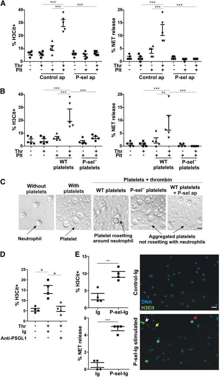
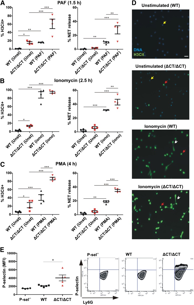
Neutrophil extracellular traps (NETs) can be released in the vasculature. In addition to trapping microbes, they promote inflammatory and thrombotic diseases. Considering that P-selectin induces prothrombotic and proinflammatory signaling, we studied the role of this selectin in NET formation. NET formation (NETosis) was induced by thrombin-activated platelets rosetting with neutrophils and was inhibited by anti-P-selectin aptamer or anti-P-selectin glycoprotein ligand-1 (PSGL-1) inhibitory antibody but was not induced by platelets from P-selectin(-/-) mice. Moreover, NETosis was also promoted by P-selectin-immunoglobulin fusion protein but not by control immunoglobulin. We isolated neutrophils from mice engineered to overproduce soluble P-selectin (P-selectin(ΔCT/ΔCT) mice). Although the levels of circulating DNA and nucleosomes (indicative of spontaneous NETosis) were normal in these mice, basal neutrophil histone citrullination and presence of P-selectin on circulating neutrophils were elevated. NET formation after stimulation with platelet activating factor, ionomycin, or phorbol 12-myristate 13-acetate was significantly enhanced, indicating that the P-selectin(ΔCT/ΔCT) neutrophils were primed for NETosis. In summary, P-selectin, cellular or soluble, through binding to PSGL-1, promotes NETosis, suggesting that this pathway is a potential therapeutic target for NET-related diseases.


Figures

Figure 1
Figure 1
P-selectin is a stimulant of NET formation. (A) Neutrophils were incubated with unstimulated or thrombin (Thr)-stimulated platelets (Plt) in the presence of P-selectin aptamer inhibitor ARC5690 (P-sel ap) or ARC5694 as control aptamer (Control ap). H3Cit+ cells and NET formation were evaluated (n = 5; ***P < .001). (B) H3Cit+ cells and NET formation were quantified in neutrophils incubated with unstimulated or thrombin-stimulated platelets from WT or P-selectin−/− mice (P-sel−/−) (n = 5; **P < .01, ***P < .001). (C) The behavior of platelets in panels A and B was visualized by differential interference contrast microscopy. The images are representative from 5 experiments (scale bar represents 5 μm). (D) NET formation was examined after incubation with unstimulated or thrombin-stimulated WT platelets in the presence of anti-PSGL-1–blocking antibody or its immunoglobulin control (Ig). The percentage of H3Cit+ neutrophils was quantified (n = 4; *P < .05). (E) Left: WT neutrophils were stimulated with P-sel-Ig or with the same class of immunoglobulin as a control (n = 4; **P < .01, ***P < .001). Right: Representative fluorescence microscopy images of control immunoglobulin (Control-Ig) or P-sel-Ig treatments showing H3Cit-negative neutrophils (yellow arrow), H3Cit+ neutrophils without NET release (red arrow), and H3Cit+ neutrophils releasing NETs (white arrowhead). Scale bar represents 20 μm.
Figure 2
Figure 2
Neutrophils of P-selectinΔCT/ΔCT mice are primed to release NETs in vitro. Neutrophils isolated from peripheral blood of WT and P-selectinΔCT/ΔCT (ΔCT/ΔCT) mice were kept unstimulated (Unst) or stimulated with PAF (A), ionomycin (iono) (B), or PMA (C). The percentage of H3Cit+ neutrophils and NET release were quantified (n = 4; *P < .05, **P < .01, ***P < .001). (D) Representative fluorescence microscopy images of unstimulated or ionomycin-stimulated cells showing H3Cit-negative neutrophils (yellow arrow), H3Cit+ neutrophils without NET release (red arrow), and H3Cit+ neutrophils releasing NETs (white arrowhead). Scale bar represents 20 μm. (E) Left: Neutrophils isolated from peripheral blood of P-selectin−/−, WT, or P-selectinΔCT/ΔCT mice were double stained with APC-conjugated anti-Ly6G and FITC-conjugated anti-P-selectin, and the mean fluorescence intensity (MFI) of P-selectin-positive cells was analyzed in the neutrophil gate (Ly6G-positive cells) by flow cytometry (n = 4-5; *P < .05). Right: Representative fluorescence-activated cell sorter plots for each genotype.

Comment in

  • Selectin' for NETs.
    Looney MR. Looney MR. Blood. 2015 Jul 9;126(2):129-30. doi: 10.1182/blood-2015-05-646729. Blood. 2015. PMID: 26160186 No abstract available.

References

    1. Brinkmann V, Reichard U, Goosmann C, et al. Neutrophil extracellular traps kill bacteria. Science. 2004;303(5663):1532–1535. - PubMed
    1. Buchanan JT, Simpson AJ, Aziz RK, et al. DNase expression allows the pathogen group A Streptococcus to escape killing in neutrophil extracellular traps. Curr Biol. 2006;16(4):396–400. - PubMed
    1. Clark SR, Ma AC, Tavener SA, et al. Platelet TLR4 activates neutrophil extracellular traps to ensnare bacteria in septic blood. Nat Med. 2007;13(4):463–469. - PubMed
    1. Xu J, Zhang X, Pelayo R, et al. Extracellular histones are major mediators of death in sepsis. Nat Med. 2009;15(11):1318–1321. - PMC - PubMed
    1. Borissoff JI, Joosen IA, Versteylen MO, et al. Elevated levels of circulating DNA and chromatin are independently associated with severe coronary atherosclerosis and a prothrombotic state. Arterioscler Thromb Vasc Biol. 2013;33(8):2032–2040. - PMC - PubMed

Publication types