Case Reports
. 2015 May 15:2015:bcr2015209368.
doi: 10.1136/bcr-2015-209368.

Metachronous multicentric giant cell tumour in a young woman

Affiliations
Case Reports

Metachronous multicentric giant cell tumour in a young woman

Raju Vaishya et al. BMJ Case Rep. .

Abstract

Multicentric giant cell tumours (GCTs) are very rare and account for less than 1% of all GCTs of bone. We report a case of a young woman with metachronous multicentric GCTs with 5 documented lesions in the same lower limb. The initial lesion started during the first trimester of pregnancy around her right pelvis, which rapidly progressed as a painful swelling with gradually restricted mobility of her right hip joint. The radiological appearance of this tumour was that of a GCT and biopsy confirmed the diagnosis. The role of positron emission tomography (PET) has been highlighted to detect occult lesions. A possible hormonal correlation for these tumours has been discussed. The patient was managed successfully by an aggressive surgical approach for knee and talar lesions, whereas repeated embolisation and denosumab injections were given to treat her pelvic lesion.


Figures

Figure 1
Figure 1
Anteroposterior radiograph of the pelvis showing a large osteolytic lesion involving the right side of the pelvis and proximal femur.
Figure 2
Figure 2
Photomicrograph (×4 magnification) of the core biopsy of right distal femur, showing numerous multinucleated giant cells evenly distributed in sheets of stromal cells, suggestive of giant cell tumour.
Figure 3
Figure 3
Lateral radiograph of the right knee, showing osteolytic lesion with intralesional calcification in the distal femur and hidden patella with large soft tissue swelling.
Figure 4
Figure 4
(A) Positron emission tomography (PET) CT scan image of the right hip showing a large pelvic tumour with increased uptake of fluorodeoxyglucose (FDG). (B) PET CT scan image of the right knee showing increased uptake of FDG. (C) PET CT scan image of the right ankle showing increased uptake of FDG.
Figure 5
Figure 5
Lateral radiograph of the right ankle showing a large lytic lesion in the talus.
Figure 6
Figure 6
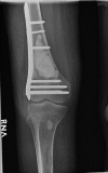
Anteroposterior radiograph of the right knee showing bone cementation in distal femur and locking plate fixation.
Figure 7
Figure 7
Follow-up radiograph of the pelvis showing diffuse calcification of the pelvic tumour.

References

    1. Manaster BJ, Doyle AJ. Giant cell tumors of bone. Radiol Clin North Am 1993;31:299–323. - PubMed
    1. Hoch B, Inwards C, Sundaram M et al. . Multicentric giant cell tumor of bone: clinicopathologic analysis of thirty cases. J Bone Joint Surg Am 2006;88:1998–2008. 10.2106/JBJS.E.01111 - DOI - PubMed
    1. Dhillon MS, Prasad P. Multicentric giant cell tumor of bone. Acta Orthop Belg 2007;73:289–99. - PubMed
    1. Xu SF, Adams B, Yu XC et al. . Denosumab and giant cell tumor of bone—a review and future management considerations. Curr Oncol 2013;20:e442–7. 10.3747/co.20.1497 - DOI - PMC - PubMed
    1. Oursler MJ, Pederson L, Fitzpatrick L et al. . Human giant cell tumors of the bone (osteoclastomas) are estrogen target cells. Proc Natl Acad Sci USA 1994;91:5227–31. 10.1073/pnas.91.12.5227 - DOI - PMC - PubMed

Publication types

MeSH terms

LinkOut - more resources