. 2015 May 15;308(10):C803-12.
doi: 10.1152/ajpcell.00223.2014. Epub 2015 Mar 11.

Endoplasmic reticulum and oxidant stress mediate nuclear factor-κB activation in the subfornical organ during angiotensin II hypertension

Affiliations

Endoplasmic reticulum and oxidant stress mediate nuclear factor-κB activation in the subfornical organ during angiotensin II hypertension

Colin N Young et al. Am J Physiol Cell Physiol. .

Abstract

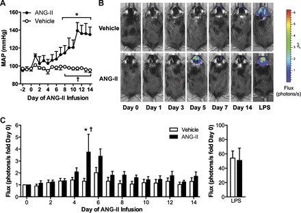
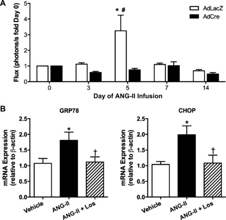
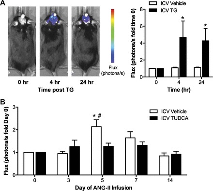
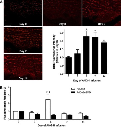
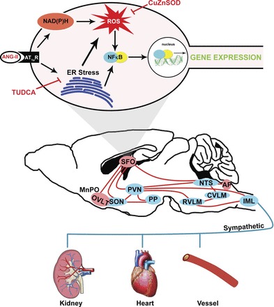
Endoplasmic reticulum (ER) stress and reactive oxygen species (ROS) generation in the brain circumventricular subfornical organ (SFO) mediate the central hypertensive actions of Angiotensin II (ANG II). However, the downstream signaling events remain unclear. Here we tested the hypothesis that angiotensin type 1a receptors (AT1aR), ER stress, and ROS induce activation of the transcription factor nuclear factor-κB (NF-κB) during ANG II-dependent hypertension. To spatiotemporally track NF-κB activity in the SFO throughout the development of ANG II-dependent hypertension, we used SFO-targeted adenoviral delivery and longitudinal bioluminescence imaging in mice. During low-dose infusion of ANG II, bioluminescence imaging revealed a prehypertensive surge in NF-κB activity in the SFO at a time point prior to a significant rise in arterial blood pressure. SFO-targeted ablation of AT1aR, inhibition of ER stress, or adenoviral scavenging of ROS in the SFO prevented the ANG II-induced increase in SFO NF-κB. These findings highlight the utility of bioluminescence imaging to longitudinally track transcription factor activation during the development of ANG II-dependent hypertension and reveal an AT1aR-, ER stress-, and ROS-dependent prehypertensive surge in NF-κB activity in the SFO. Furthermore, the increase in NF-κB activity before a rise in arterial blood pressure suggests a causal role for SFO NF-κB in the development of ANG II-dependent hypertension.

Keywords: NF-κB, ER stress; angiotensin II; central nervous system; hypertension.


Figures

Fig. 1.
Fig. 1.
Slow-pressor angiotensin II (ANG II) infusion evokes a prehypertensive surge of nuclear factor-κB (NF-κB) in the subfornical organ (SFO). A: radiotelemetry measurements of mean arterial blood pressure (MAP) during infusion of vehicle (BSA) or ANG II. B: original bioluminescence images of NF-κB-dependent photon emission during infusion of vehicle or ANG II. The areas of high photon emission (flux) are displayed in red, and the areas of low photon emission are displayed in blue. C: summary of temporal changes in NF-κB activity. Changes in NF-κB activity in response to systemic lipopolysaccharide (LPS) at the end of the studies are shown on the right; note the different scale; n = 9–10/group. *P < 0.05 vs. day 0; †P < 0.05 vs. vehicle.
Fig. 2.
Fig. 2.
Angiotensin type 1a receptors (AT1aR) mediate ANG II-induced SFO NF-κB activation and endoplasmic reticulum (ER) stress. A: summary of temporal changes in NF-κB activity at key time points during ANG II infusion in AT1aRfl/fl mice with SFO-targeted adenoviral delivery of Cre-recombinase (AdCre) or control, AdLacZ; n = 5/group. *P < 0.05 vs. day 0; #P < 0.05 vs. AdCre. B: GRP78 (left) and CHOP (right) mRNA expression in Neuro2A cells treated with vehicle, ANG II, or ANG II plus losartan (Los). *P < 0.05 vs. vehicle; †P < 0.05 vs. ANG II.
Fig. 3.
Fig. 3.
ER stress mediates ANG II-induced NF-κB activation in the SFO. A: original bioluminescence images of NF-κB-dependent photon emission during intracerebroventricular (icv) infusion of vehicle or the ER stress inducer thapsigargin (TG). Group summary data are shown on the right; n = 7–8/group. *P < 0.05 vs. time 0. B: summary of temporal changes in NF-κB activity during ANG II infusion in mice treated daily with icv vehicle or the ER stress inhibitor tauroursodeoxycholic acid (TUDCA); n = 7–8/group. *P < 0.05 vs. day 0; #P < 0.05 vs. ICV TUDCA.
Fig. 4.
Fig. 4.
ANG II-induced activation of NF-κB in the SFO is mediated by oxidative stress. A: representative dihydroethidium (DHE) fluorescence images of the SFO from untreated mice and at key time points during ANG II infusion. Scale bar = 50 μm. B: summary of temporal changes in NF-κB activity at key time points during ANG II infusion in mice with SFO-targeted adenoviral delivery of cytoplasmic superoxide dismutase (AdCuZnSOD) or control, AdLacZ; n = 8–10/group. *P < 0.05 vs. day 0; #P < 0.05 vs. AdCuZnSOD.
Fig. 5.
Fig. 5.
ANG II does not cause cell death or activate activator protein-1 (AP-1) in the SFO. A: representative terminal deoxynucleotidyl transferase dUTP nick end labeling (TUNEL) staining (green) in SFO sections from untreated (day 0) and ANG II-infused mice (day 14). A positive control section from the intestine is shown on the right. DAPI staining is shown in blue; n = 3. Scale bar = 100 μm. B: summary of temporal changes in SFO AP-1-dependent photon emission during ANG II infusion; n = 6–8/group.
Fig. 6.
Fig. 6.
Schematic illustrating the central mechanisms within the SFO that may mediate ANG II-dependent hypertension. ANG II, via AT1aR, induces ER stress and NADPH oxidase-dependent reactive oxygen species (ROS) production in the SFO. ER stress and oxidant signaling lead to activation of NF-κB, subsequent changes in gene expression, and downstream stimulation of sympathoexcitatory circuits. MnPO, median preoptic nucleus; OVLT, organum vasculosum lamina terminalis; SON, supraoptic nucleus; PVN, paraventricular nucleus; PP, posterior pituitary; RVLM, rostral ventral lateral medulla; CVLM, caudal ventral lateral medulla; NTS, nucleus tractus solitarii; AP, area postrema; IML, intermediolateral column of the spinal cord.

References

    1. Blume A, Herdegen T, Unger T. Angiotensin peptides and inducible transcription factors. J Mol Med 77: 339–357, 1999. - PubMed
    1. Brasier AR, Jamaluddin M, Han Y, Patterson C, Runge MS. Angiotensin II induces gene transcription through cell-type-dependent effects on the nuclear factor-kB (NF-kB) transcription factor. 212: 155–169, 2000. - PubMed
    1. Burmeister MA, Young CN, Braga VA, Butler SD, Sharma RV, Davisson RL. In vivo bioluminescence imaging reveals redox-regulated activator protein-1 activation in paraventricular nucleus of mice with renovascular hypertension. Hypertension 57: 289–297, 2011. - PMC - PubMed
    1. Butz GM, Davisson RL. Chronic blood pressure recording in pregnant mice by radiotelemetry. Physiol Genomics 2001.
    1. Cao X, Peterson JR, Wang G, Anrather J, Young CN, Guruju MR, Burmeister MA, Iadecola C, Davisson RL. Angiotensin II-dependent hypertension requires cyclooxygenase 1-derived prostaglandin E2 and EP1 receptor signaling in the subfornical organ of the brain. Hypertension 59: 869–876, 2012. - PMC - PubMed

Publication types

MeSH terms

LinkOut - more resources