. 2015 Jul 20;6(20):18050-65.
doi: 10.18632/oncotarget.3777.

XB130 translocation to microfilamentous structures mediates NNK-induced migration of human bronchial epithelial cells

Affiliations

XB130 translocation to microfilamentous structures mediates NNK-induced migration of human bronchial epithelial cells

Qifei Wu et al. Oncotarget. .

Abstract

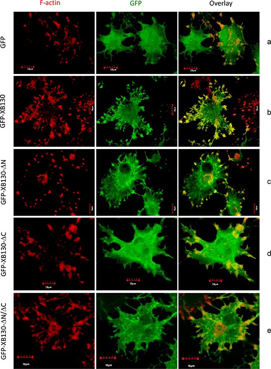
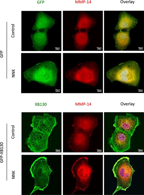
Cigarette smoking contributes to the pathogenesis of chronic obstructive pulmonary disease and lung cancer. Nicotine-derived nitrosamine ketone (NNK) is the most potent carcinogen among cigarette smoking components, and is known to enhance migration of cancer cells. However, the effect of NNK on normal human bronchial epithelial cells is not well studied. XB130 is a member of actin filament associated protein family and is involved in cell morphology changes, cytoskeletal rearrangement and outgrowth formation, as well as cell migration. We hypothesized that XB130 mediates NNK-induced migration of normal human bronchial epithelial cells. Our results showed that, after NNK stimulation, XB130 was translocated to the cell periphery and enriched in cell motility-associated structures, such as lamellipodia, in normal human bronchial epithelial BEAS2B cells. Moreover, overexpression of XB130 significantly enhanced NNK-induced migration, which requires both the N- and C-termini of XB130. Overexpression of XB130 enhanced NNK-induced protein tyrosine phosphorylation and promoted matrix metalloproteinase-14 translocation to cell motility-associated cellular structures after NNK stimulation. XB130-mediated NNK-induced cell migration may contribute to airway epithelial repair; however, it may also be involved in cigarette smoking-related chronic obstructive pulmonary disease and lung cancer.

Keywords: F-actin association; airway epithelial cell migration; cortactin; intracellular signal transduction.


Figures

Figure 1
Figure 1. NNK-induces translocation of XB130 to actin-rich punctate and lamellipodia at the periphery of BEAS2B cells
A. NNK-stimulation induced translocation of endogenous XB130 from cytoplasm to actin-rich small dots and lamellipodia at the periphery of the cells. BEAS2B cells were stimulated with or without 0.1 μM NNK for 30 min. Cells were immunostained with anti-human XB130 antibody and Alexa Fluor 594 labeled secondary antibody (red), and co-stained with Alexa Fluor 488 phalloidin (green). B. Overexpression of GFP-XB130 increased association of XB130 with actin stress fibers under control condition, and enhanced NNK-induced translocation of XB130 to actin-rich cellular structures. BEAS2B cells stably transfected with GFP-XB130 were treated with or with 0.1 μM NNK for 30 min. F-actin was stained with Alexa Fluor 594 phalloidin (red). Scale bar is 10 μm.
Figure 2
Figure 2. NNK induces association of XB130 with actin cytoskeletal structures. BEAS2B cells were stably transfected with GFP-XB130 or GFP alone
A. GFP-XB130, but not GFP alone, is partially retained with actin cytoskeletal structures. After NNK stimulation (0.1 μM for 30 min), cells were treated with 0.2% Triton X-100 buffer for 5 min to remove cytoplasm. F-actin was stained with Alexa Fluor 594 phalloidin (red). Scale bar is 10 μm. B. XB130 is in the membranous cytoskeleton. GFP-XB130 cells were stimulated with 0.1 μM NNK for 30 min. Cell lysates were differentially centrifuged to collect the crude cytoskeleton fraction (10,000 g, pellet), cytoplasm fraction (100,000 g, supernatant), and membranous cytoskeleton fraction (100,000 g, pellet). XB130 was found mainly in the cytoplasm and membranous cytoskeletal fraction (see DNase I- group). DNase (0.5 mg/ml) treatment resulted in the loss of both actin and XB130 from the membrane cytoskeleton fraction of BEAS2B cell lysates (see DNase I+ group).
Figure 3
Figure 3. Overexpression of XB130 enhances BEAS2B cell migration. BEAS2B cells were stably transfected with GFP-XB130 or GFP alone
A. Wound healing assays showed GFP-XB130 transfected cells showed increased cell migration as compared to GFP only transfected cells (n = 3, *P < 0.05). B. NNK-induced rapid translocation of XB130. BEAS2B cells stably transfected with GFP-XB130 or GFP alone were seeded in 4-well chamber slides. Cell images were captured before and after NNK treatment (0.1 μM for 15 min at 37°C, 5% CO2) using a Zeiss Apotome at 63x oil objective lens. Live cell-imaging shows that the distribution of GFP alone was not affected by NNK stimulation. By contrast, GFP-XB130 was seen at the front edge of the cell, which was further enhanced immediately after adding NNK (indicated by arrow head). GFP-XB130 also formed punctate structures inside of cells. Shown are representative images taken every minute after NNK treatment for a total of 3 min. Scale bar is 20 μm.
Figure 4
Figure 4. Overexpression of XB130 increased chemotactic cell migration
A. GFP-XB130 enhanced NNK-induced cell migration. BEAS2B cells were seeded in QCM™ 24-well transwell chamber for chemotaxis cell migration assay. Cells were stimulated with 10% FBS together with or without 0.1 μM NNK for 16 h. Migration of GFP-XB130 transfected cells was significantly fast than that of GFP alone cells and Control cells (#: P < 0.05 vs. Control or GFP alone group). GFP-XB130 significantly increased NNK-induced cell migration (n = 3, *: P < 0.05, Compared between with and without NNK stimulation on GFP-XB130 groups). B. A schematic diagram representing the wild type full length XB130 protein and various deletion mutants used to develop stable transfection cell lines. C. The N- and C-termini of XB130 are required for mediating NNK-induced cell migration. Cells stably transfected with GFP-XB130 or its deletion mutants were stimulated with 10% FBS with 0.1 μM NNK for 16 h for chemotaxis cell migration assay. Deletion of N- and/or C-terminus of XB130 reduced NNK-induced cell migration (n = 3, *P < 0.05). UR: a unique region that is seen only in XB130, but not in other actin filament associate protein family members.
Figure 5
Figure 5. The C-terminus of XB130 is required for NNK-induced redistribution of XB130 to actin-rich aggregates
Cells stably transfected with GFP-XB130 or its GFP-labeled mutants (green) were stimulated with 0.1 μM NNK for 30 min followed by 2 μM cytochalasin D treatment at 37°C for 1 h to inhibit F-actin polymerization. F-actin was stained with Texas-red conjugated phalloidin (red). Representative images show that deletion of the C-terminus or both C- and N-termini of XB130 prevented co-localization of GFP-XB130 with F-actin aggregates. Scale bar is 10 μm.
Figure 6
Figure 6. NNK treatment increased tyrosine phosphorylation of XB130
A. GFP-XB130 enhanced NNK-induced protein tyrosine phosphorylation. BEAS2B cells stably transfected with GFP-XB130 or GFP vector alone were stimulated with 0.1 μM NNK for up to 60 min. Immunoblotting with anti-phosphotyrosine antibody (p-Tyr) shows that NNK increased multiple protein tyrosine phosphorylation, which was further enhanced by XB130 overexpression. B and C. NNK enhanced XB130 tyrosine phosphorylation. GFP-XB130 cells were stimulated with 0.1 μM NNK for up to 30 min. Whole cell lysates were immunoprecipitated with anti-XB130 antibody B. or anti-phosphotyrosine antibody C. and immunoblotted with antibodies indicated. NNK enhanced XB130 tyrosine phosphorylation after 15 min. D. NNK-induced XB130 tyrosine phosphorylation at the lamellipodia. Cells stably transfected with GFP-XB130 or GFP vector alone were stimulated with 0.1 μM NNK for 30 min. Immunostaining for anti-phosphotyrosine antibody using Alexa Fluor 594 labeled secondary antibody (red) and detection of GFP signals show that NNK stimulation did not induced translation of GFP alone (top panel). By contrast, NNK increased protein tyrosine phosphorylation and co-localization of GFP-XB130 and phosphotyrosine at the cell periphery (see overlay and inset). Scale bar is 20 μm.
Figure 7
Figure 7. XB130/cortactin interaction is enhanced by NNK treatment in BEAS2B cells
A. BEAS2B cells stably expressing GFP-XB130 or GFP alone were treated with 0.1 μM NNK for 30 min. Cells were stained with anti-cortactinantibody and Alexa Fluor 594 labeled secondary antibody (red), and Alexa Fluor 350 conjugated phalloidin (blue) for F-actin. In comparison with GFP alone transfected cells, GFP-XB130 enhanced NNK-induced translation of cortactin and colocalized with cortactin within actin-rich structures at the cell periphery. Scale bar is 50 μm. B. Co-immunoprecipitation between XB130 and cortactin was increased by NNK stimulation. GFP-XB130 stably transfected BEAS2B cells were treated with 0.1 μM NNK up to 30 min. Cell lysates were co-immunoprecipitated anti-cortactin or anti-XB130 antibody and immunoblots of XB130 and cortactin show that the interaction between XB130 and cortactin was enhanced by NNK stimulation.
Figure 8
Figure 8. Over-expression of XB130 promoted MMP-14 translocation to actin-rich cellular structures
BEAS2B cells stably transfected with GFP-XB130 or GFP alone were stimulated with 0.1 μM NNK for 30 min. Overexpression of XB130 enhanced NNK-induced MMP-14 (red) translocationto lamellipodia and ruffles on the cell peripheral membrane. Scale bar is 10 μm.

Similar articles

Cited by

References

    1. Park H, Shin JW, Park SG, Kim W. Microbial communities in the upper respiratory tract of patients with asthma and chronic obstructive pulmonary disease. PLoS One. 2014;9:e109710. - PMC - PubMed
    1. Jemal A, Murray T, Samuels A, Ghafoor A, Ward E, Thun MJ. Cancer statistics, 2003. CA Cancer J Clin. 2003;53:5–26. - PubMed
    1. Decramer M, Janssens W, Miravitlles M. Chronic obstructive pulmonary disease. Lancet. 2012;379:1341–1351. - PMC - PubMed
    1. West KA, Brognard J, Clark AS, Linnoila IR, Yang X, Swain SM, Harris C, Belinsky S, Dennis PA. Rapid Akt activation by nicotine and a tobacco carcinogen modulates the phenotype of normal human airway epithelial cells. J Clin Invest. 2003;111:81–90. - PMC - PubMed
    1. West KA, Linnoila IR, Belinsky SA, Harris CC, Dennis PA. Tobacco carcinogen-induced cellular transformation increases activation of the phosphatidylinositol 3′-kinase/Akt pathway in vitro and in vivo. Cancer Res. 2004;64:446–451. - PubMed

Publication types

MeSH terms

Substances

LinkOut - more resources