. 2015 Jun;53(6):366-76.
doi: 10.1002/dvg.22859. Epub 2015 May 30.

A Hoxa13:Cre mouse strain for conditional gene manipulation in developing limb, hindgut, and urogenital system

Affiliations

A Hoxa13:Cre mouse strain for conditional gene manipulation in developing limb, hindgut, and urogenital system

Martina Scotti et al. Genesis. 2015 Jun.

Abstract

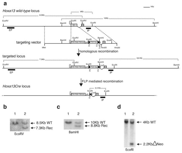
The developing limb is a useful model for studying organogenesis and developmental processes. Although Cre alleles exist for conditional loss- or gain-of-function in limbs, Cre alleles targeting specific limb subdomains are desirable. Here we report on the generation of the Hoxa13:Cre line, in which the Cre gene is inserted in the endogenous Hoxa13 gene. We provide evidence that the Cre is active in embryonic tissues/regions where the endogenous Hoxa13 gene is expressed. Our results show that cells expressing Hoxa13 in developing limb buds contribute to the entire autopod (hand/feet) skeleton and validate Hoxa13 as a distal limb marker as far as the skeleton is concerned. In contrast, in the limb musculature, Cre-based fate mapping shows that almost all muscle masses of the zeugopod (forearm) and part of the triceps contain Hoxa13-expressing cells and/or their descendants. Besides the limb, the activity of the Cre is detectable in the urogenital system and the hindgut, primarily in the epithelium and smooth muscles. Together our data show that the Hoxa13:Cre allele is a useful tool for conditional gene manipulation in the urogenital system, posterior digestive tract, autopod and part of the limb musculature.

Keywords: fate specification; genetics; gut; limb/wing/appendage; mammal; muscle; organism; organogenesis; process; reproductive; skeletal; tissue.


Figures

FIG. 1
FIG. 1
Generation of the Hoxa13:Cre mouse line. (a) Targeting of the endogenous Hoxa13 locus. (Top) Wild-type Hoxa13 locus. The targeting vector is shown below and dotted lines indicate the position of the homologous arms. (Middle) Scheme of the targeted locus after homologous recombination in ES cells. The position of the internal (IP) and external probes (EP) and restriction sites used for Southern analysis are indicated. (Bottom) The targeted locus after FLP-mediated deletion of the PGK-Neo selection cassette. (b) Southern blots of ES clones using the external probe and (c) the internal probe to detect the targeted allele (lane 2). (d) Southern blot analysis of wild type (lane 1) and heterozygous (lane 2) mice after in vivo FLP-mediated deletion.
FIG. 2
FIG. 2
Comparison between Hoxa13 expression and the localization of Hoxa13lin+ cells in the developing embryo. (a, d, g, j, m, p) Whole-mount in situ hybridization of wild-type embryos with the Hoxa13 RNA probe. (b, c, e, f, h, i, k, l, n, o) Whole-mount X-gal staining of Hoxa13:Cre/+; Rosa26R/+ embryos. Black arrowhead in (a) indicates Hoxa13 expression domain in the developing limb bud. Dotted lines in (b) demarcate the forelimb bud. (o) X-gal staining of the gastro-intestinal track. TB: tail bud; UC: umbilical cord; US: urogenital sinus; NT: neural tube; s: stomach; ce: caecum; co: colon; re: rectum.
FIG. 3
FIG. 3
Contribution of Hoxa13lin+ cells to the urogenital system. (a–y) Co-immunostaining for GFP and alpha smooth muscle actin (SMA) on cryosections of E18.5 Hoxa13Cre/+; mT/mG fetuses. (a–e) Frontal section showing the presence Hoxa13lin+ cells in ureter but not to the adrenal (suprarenal) gland (ag) nor kidney (K). (f–j) Higher magnification of the proximal region of the ureter. Note the absence of Hoxa13lin+ cells in the kidney-ureter junction. (k–o) Transverse section through the ureter. (p–t) Frontal sections of the bladder. Nuclei are stained with DAPI in panels a, f, k, p, u. mT (red fluorescence) is shown as a control of GFP-negative cells. ad: adventitia; ag: adrenal gland; ct: connective tissue; ilm: inner longitudinal muscular layer; k: kidney; lu: lumen; ocm: outer circular muscular layer; sm: smooth muscle; u: ureter; ur: urethra; uro: urothelium; ut: uterus; vc: vesicular cavity.
FIG. 4
FIG. 4
Fate map of Hoxa13-expressing cells in the developing hindgut. (a–j) Transverse section of E18.5 developing colon isolated from Hoxa13Cre/+; mT-mG/+ embryos. (f–j) High magnification of the dotted square in panels a–e. Hoxa13lin+ cells (green, mG-expressing cells) are present in the serosa (se), longitudinal fibers (lf), and circular fibers (cf) (muscular coat), mucous coat (mc) and the epithelium lining the lumen (ep). en: enteric plexus.
FIG. 5
FIG. 5
Cre reporter expression in the developing forelimb muscles. (a) Whole-mount X-gal staining of Hoxa13:Cre/1;Rosa26R/1 forelimb buds from e12.5 to postnatal day 2 (P2). The Cre reporter expression is observed in the entire autopod as well as in more proximal regions of the developing limb, where its expression suggests expression in the developing limb musculature and/or associated connective tissue. Scale bars5100 mm. (b–g) Co-immunostaining for GFP and fast skeletal Myosin on transverse section of the Hoxa13:Cre/1; mT-mG/1 zeupogod at E14.5. View of the flexor digitorium sublimis (b–d) and extensor digitorium communis (e–g). Note that there are few scattered Hoxa13lin1 cells that do not express fast skeletal Myosin (white arrows). (h–m) Co-immunostaining for GFP and Tcf4. (h–j) View of the extensor digitorium communis and lateralis. (k–m) high magnification of the dotted rectangles in panels h to j. Yellow arrowheads point to example of cells expressing Tcf4 but not GFP (i.e. cells that are not Hoxa13lin+ cells). White arrowheads point to the few Hoxa13lin+ cells that express Tcf4, though at lower level compared with the other Tcf4-expressing cells. (n–p) Transverse section of Hoxa13:Cre/+; mT-mG/ + zeupogod at E14.5 showing the overall distribution of Hoxa13lin positive cells (mG-expressing cells in green) and Hoxa13lin negative cells (mT-expressing cells in red). Note the nonuniform distribution of mG-expressing cells, in particular in the Extensor Carpi Ulnaris (dotted rectangle), suggesting a clonal contribution of Hoxa13lin+ cells to the various muscles.
FIG. 6
FIG. 6
Differential contribution of Hoxa13lin+ cells to the limb muscles. Expression pattern of GFP and fast skeletal Myosin (my-32) on transverse sections of Hoxa13:Cre/+; mT-mG/+ forelimb bud at E14.5 (a–i) and E18.5 (j–r). Transverse sections through the distal (a–c; j–l) and proximal part (d–f; m–o) of the zeugopod and through the stylopod (g–i; p–r). For muscle nomenclature, see Table 1. R: radius; U: ulna; H: humerus; dor: dorsal; ven: ventral; ant: anterior; post: posterior.
FIG. 7
FIG. 7
Hoxa13-expressing cells give rise to all bones of the autopod and mark the boundary between the wrist and the zeugopod. (a–c) Sagittal section through a E14.5 Hoxa13:Cre/+;mT-mG/+ forelimb showing the overall distribution of Hoxa13lin positive cells (mG-expressing cells; green) and Hoxa13lin negative cells (mT-expressing cells; red). Distal is on the left and anterior is up. (d–f) Sagittal section at the autopod-zeugopod boundary of E18.5 Hoxa13:Cre/+;mT-mG/+ forelimb. Hoxa13lin+ cells are visualized by direct detection of GFP fluorescence from the Cre reported allele (green) and immunostaining for my-32 (red) marks muscles. Note that all carpal bones are GFP+ and only few GFP+ cells are present in the ulna head (U). (g–i) high magnification of the ulna head (U) and ulnare (ul) showing few Hoxa13lin negative cells in the ulnare (mT-expressing cells; red) while a small cluster of Hoxa13lin+ cells (green) are present in the ulna head (U). Nuclei are stained with DAPI (blue). R: radius; U: ulna; ra: radiale; i: intermedium; ul: ulnare.

References

    1. Copeland NG, Jenkins NA, Court DL. Recombineering: A powerful new tool for mouse functional genomics. Nat Rev Genet. 2001;2:769–779. - PubMed
    1. Dolle P, Duboule D. Two gene members of the murine HOX-5 complex show regional and cell-type specific expression in developing limbs and gonads. EMBO J. 1989;8:1507–1515. - PMC - PubMed
    1. Farley FW, Soriano P, Steffen LS, Dymecki SM. Widespread recombinase expression using FLPeR (flipper) mice. Genesis. 2000;28:106–110. - PubMed
    1. Fromental-Ramain C, Warot X, Messadecq N, LeMeur M, Dolle P, Chambon P. Hoxa-13 and Hoxd-13 play a crucial role in the patterning of the limb auto-pod. Development. 1996;122:2997–3011. - PubMed
    1. Haack H, Gruss P. The establishment of murine Hox-1 expression domains during patterning of the limb. Dev Biol. 1993;157:410–422. - PubMed

Publication types

MeSH terms

LinkOut - more resources