Review
. 2015 Oct 7;589(20 Pt A):2914-22.
doi: 10.1016/j.febslet.2015.05.016. Epub 2015 May 14.

A brief review of nucleosome structure

Affiliations
Review

A brief review of nucleosome structure

Amber R Cutter et al. FEBS Lett. .

Abstract

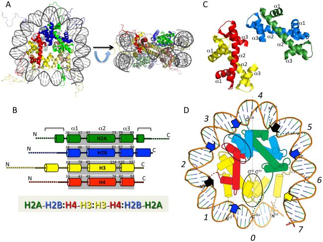
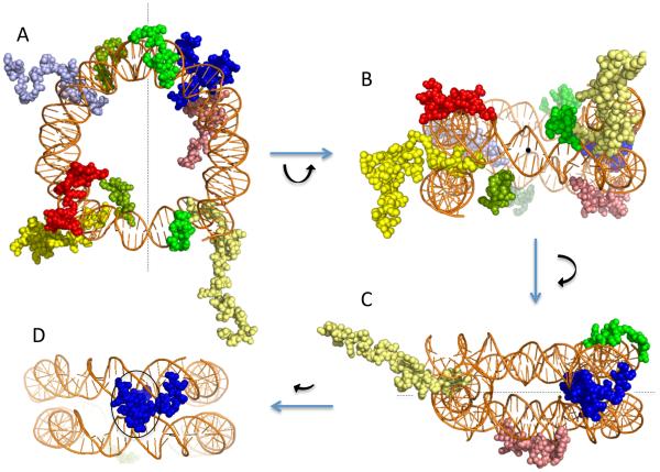
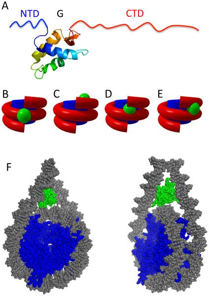
The nucleosomal subunit organization of chromatin provides a multitude of functions. Nucleosomes elicit an initial ∼7-fold linear compaction of genomic DNA. They provide a critical mechanism for stable repression of genes and other DNA-dependent activities by restricting binding of trans-acting factors to cognate DNA sequences. Conversely they are engineered to be nearly meta-stable and disassembled (and reassembled) in a facile manner to allow rapid access to the underlying DNA during processes such as transcription, replication and DNA repair. Nucleosomes protect the genome from DNA damaging agents and provide a lattice onto which a myriad of epigenetic signals are deposited. Moreover, vast strings of nucleosomes provide a framework for assembly of the chromatin fiber and higher-order chromatin structures. Thus, in order to provide a foundation for understanding these functions, we present a review of the basic elements of nucleosome structure and stability, including the association of linker histones.

Keywords: Chromatin structure; Histone; Nucleosome structure.


Figures

Fig. 1
Fig. 1. Structural Details of a Nucleosome Core
A. Model of a nucleosome core (PDB: 1KX5, ref: [20]). Shown are a view down the superhelical axis, and a view rotated 90°about a horizontal axis, as shown, looking down the dyad axis of the nucleosome. H2A, green, H2B, blue, H3, yellow, H4, red. Proteins in lower half of nucleosome are lighter in color. B. Top: Schematic showing secondary structure of the core histone proteins, with α-helices represented by columns. Dashed lines indicate approximate residues within ‘tail’ domains; shaded boxes indicate the 3-helix histone fold domains within each protein, with first and last residues within α1, α2 and α3 helices indicated. Additional helices outside the histone fold domain are indicated by brackets, Bottom: Linear representation of primary contacts between the core histone proteins in the nucleosome core. Core histone dimerization partners are separated by dashes; dimer-dimer interactions via 4-helix bundles are indicated by colons. C. H2A-H2B (green/blue) and H3-H4 (yellow/red) histone fold domain dimers. α1, α2 and α3 helices indicated, corresponding to B. D. Schematic showing one-half of nucleosome core, looking down the DNA superhelix axis. Superhelix sites are indicated by italicized numerals; 4-helix bundles between H3:H3 and H4:H2B are highlighted by ovals; blue and black arrows indicate paired loop and paired-end-of-helix DNA interaction sites. Yellow arrow indicates site of interaction centered on the N-helix in H3. Note, a small amount of DNA and H3 from the non-depicted half of the nucleosome core are shown for clarity, lighter in color.
Fig. 2
Fig. 2. Core histone tail domains and nucleosome DNA
Several orientations with only tail domains and nucleosome DNA are shown from an X-ray crystal structure model of a nucleosome core (PDB: 1KX5, ref: [20]). A. View down the nucleosome superhelix axis. B. View rotated 90°, looking down the nucleosome dyad axis. C. View rotated 90°about vertical axis looking orthogonal to dyad axis, showing tails of H3 (yellow) and H2B (blue) exiting through the superhelical gyres while the H4 (red) and H2A (green) tails exit over/under the superhelix. D. View with additional 30°rotation about vertical axis, showing only H2B tail exiting through aligned minor grooves (black oval). Protein colors as in Fig. 1. Nucleosome dyad axis is indicated (dotted line).
Fig. 3
Fig. 3. Model of H1 binding to nucleosome
A. Domain structure of metazoan linker histone showing the 25-35 residue ‘unstructured’ N-terminal domain (NTD), ~80 residue globular domain (G), and the 100-120 residue ‘unstructured’ C-terminal domain (CTD). Model of the globular domain from ref [91]. B-E. Simplified models showing approximate sites for interaction of H1 globular domain with the nucleosome from refs. [81,112,113,115], respectively. F. Three-contact model for linker histone globular domain binding within a nucleosome. Two views of a nucleosome with proposed location of a linker histone globular domain (green) are shown, with DNA and core histones colored grey and blue, respectively. Approximately 20 additional bp of DNA are depicted beyond the boundary of the nucleosome core region.

References

    1. Luger K, Mader AW, Richmond RK, Sargent DF, Richmond TJ. Crystal structure of the nucleosome core particle at 2.8 A resolution. Nature. 1997;389:251–60. - PubMed
    1. White CL, Suto RK, Luger K. Structure of the yeast nucleosome core particle reveals fundamental changes in internucleosome interactions. EMBO J. 2001;20:5207–18. - PMC - PubMed
    1. Richmond TJ, Finch JT, Rushton B, Rhodes D, Klug A. Structure of the nucleosome core particle at 7 A resolution. Nature. 1984;311:532–7. - PubMed
    1. van Holde KE. Chromatin. Springer Verlag; New York: 1989.
    1. Arents G, Burlingame RW, Wang BC, Love WE, Moudrianakis EN. The nucleosomal core histone octamer at 3.1 A resolution: a tripartite protein assembly and a left-handed superhelix. Proc. Natl. Acad. Sci. USA. 1991;88:10148–52. - PMC - PubMed

Publication types