. 2015 Aug;124(1):13-22.
doi: 10.1007/s11060-015-1807-0. Epub 2015 May 17.

Radiosensitivity enhancement of radioresistant glioblastoma by epidermal growth factor receptor antibody-conjugated iron-oxide nanoparticles

Affiliations

Radiosensitivity enhancement of radioresistant glioblastoma by epidermal growth factor receptor antibody-conjugated iron-oxide nanoparticles

Alexandros Bouras et al. J Neurooncol. 2015 Aug.

Abstract

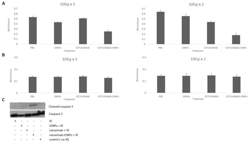
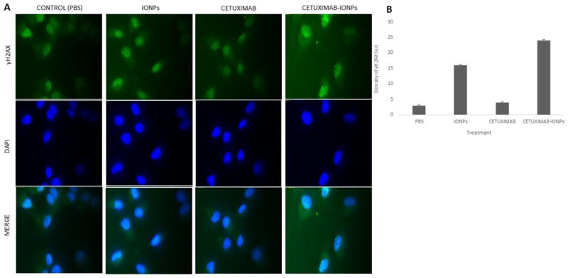
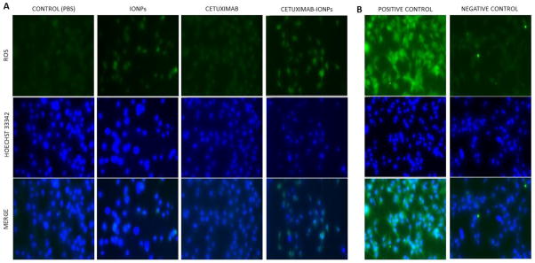
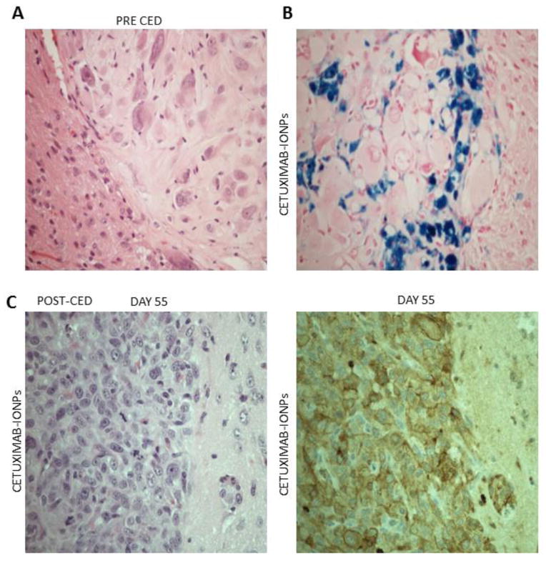
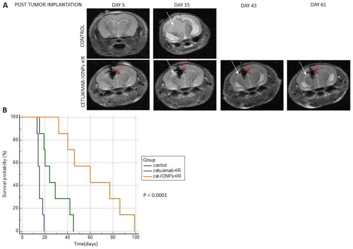
The epidermal growth factor receptor deletion variant EGFRvIII is known to be expressed in a subset of patients with glioblastoma (GBM) tumors that enhances tumorigenicity and also accounts for radiation and chemotherapy resistance. Targeting the EGFRvIII deletion mutant may lead to improved GBM therapy and better patient prognosis. Multifunctional magnetic nanoparticles serve as a potential clinical tool that can provide cancer cell targeted drug delivery, imaging, and therapy. Our previous studies have shown that an EGFRvIII-specific antibody and cetuximab (an EGFR- and EGFRvIII-specific antibody), when bioconjugated to IONPs (EGFRvIII-IONPs or cetuximab-IONPs respectively), can simultaneously provide sensitive cancer cell detection by magnetic resonance imaging (MRI) and targeted therapy of experimental GBM. In this study, we investigated whether cetuximab-IONPs can additionally allow for the radiosensitivity enhancement of GBM. Cetuximab-IONPs were used in combination with single (10 Gy × 1) or multiple fractions (10 Gy × 2) of ionizing radiation (IR) for radiosensitization of EGFRvIII-overexpressing human GBM cells in vitro and in vivo after convection-enhanced delivery (CED). A significant GBM antitumor effect was observed in vitro after treatment with cetuximab-IONPs and subsequent single or fractionated IR. A significant increase in overall survival of nude mice implanted with human GBM xenografts was found after treatment by cetuximab-IONP CED and subsequent fractionated IR. Increased DNA double strands breaks (DSBs), as well as increased reactive oxygen species (ROS) formation, were felt to represent the mediators of the observed radiosensitization effect with the combination therapy of IR and cetuximab-IONPs treatment.


Conflict of interest statement

Conflict of Interest

The authors declare no conflict of interest.

Figures

Fig. 1
Fig. 1
a & b Cell proliferation analysis (MTT Assay) of U87MGEGFRvIII cells (5 × 10³ cells/well) (a) and Normal Human Astrocytes (5 × 10³ cells/well) (b) after treatment with control (PBS), IONPs (0.3mg/ml), cetuximab (0.3mg/ml) and cetuximab-IONPs (0.3mg/ml) in combination with IR (p<0.05). C. Western blot analysis for expression of cleaved caspase 3 and caspase 3 of U87MGEGFRvIII cells after treatment with control (PBS), IONPs (0.3mg/ml), cetuximab (0.3mg/ml) and cetuximab-IONPs (0.3mg/ml) in combination with IR.
Fig. 2
Fig. 2
a Immunofluorescent staining of U87MGEGFRvIII cells (20 × 103/well) after treatment with control (PBS), IONPs (0.3mg/ml), cetuximab (0.3mg/ml), and cetuximab-IONPs (0.3mg/ml) and subsequent single IR dose of 2Gy 24 h post treatment. Fixation of cells 30 minutes post IR. Green: anti-γH2AX, Blue: DAPI. b Semi-automated density of γH2AX foci (number of foci/square inch of nucleus) for each treatment (p<0.05). Fixation of cells 30 minutes post IR
Fig. 3
Fig. 3
a ROS detection in live U87MGEGFRvIII cells (20 × 103/well) after treatment with control (PBS), IONPs (0.3mg/ml), cetuximab (0.3mg/ml) and cetuximab-IONPs (0.3mg/ml) and subsequent single IR dose of 10Gy 24 h post-treatment. Cells were stained 3 h post IR for ROS detection with 5-(and-6)-carboxy-2′,7′ dichlorodihydrofluorescein diacetate (carboxy-H2DCFDA), a marker for ROS detection in live cells. Green: ROS, Blue: Hoechst 33342. b. Left: Induction of ROS in U87MGEGFRvIII cells by tert-butyl hydroperoxide (TBHP), an inducer of ROS production (positive control). Right: U87MGEGFRvIII stained for baseline ROS expression with 5-(and-6)-carboxy-2′,7′ dichlorodihydrofluorescein diacetate (carboxy-H2DCFDA), a marker for ROS detection in live cells (negative control). Green: ROS, Blue: Hoechst 33342
Fig. 4
Fig. 4
a Hematoxylin & Eosin (H&E) staining of intracranial human U87MGEGFRvIII GBM xenograft in athymic nude mouse confirms xenograft formation prior to CED (magnification 40x). b Prussian Blue staining of athymic nude mouse brain section showing intratumoral and peritumoral distribution of bioconjugated cetuximab-IONPs after CED (magnification 40x). c H&E staining (left) and immunostaining for EGFRvIII (right) of an athymic nude mouse which underwent intracranial human U87MGEGFRvIII GBM xenograft implantation and subsequent CED of bioconjugated cetuximab-IONPs (magnification 40x)
Fig. 5
Fig. 5
a Top: T2-weighted MRI of a control mouse implanted with EGFRvIII-expressing orthotopic human GBM xenograft (U87MGEGFRvIII) showing a hyperintense xenograft (white arrows) post tumor implantation. Bottom: Serial T2-weighted MRI of a mouse which underwent implantation of an EGFRvIII-expressing orthotopic human GBM xenograft (U87MGEGFRvIII) and subsequent CED of cetuximab-IONPs. Hypointense MRI signal drop after CED of cetuximab-IONPs (red arrows) and hyperintense EGFRvIII-expressing human GBM xenograft (white arrows). b Kaplan Meier survival curve comparison of athymic nude mice after intracranial implantation of U87MG EGFRvIII cells (2 × 10⁵/mouse) receiving no treatment (control group) or combination treatment by CED of cetuximab (0.3mg/ml) or cetuximab-IONPs (0.3mg/ml) and subsequent IR (10 Gy x 2)

References

    1. Braun S, Oppermann H, Mueller A, Renner C, Hovhannisyan A, Baran-Schmidt R, Gebhardt R, Hipkiss A, Thiery J, Meixensberger J, Gaunitz F. Hedgehog signaling in glioblastoma multiforme. Cancer Biol Ther. 2012;13(7):487–495. doi: 10.4161/cbt.19591. - DOI - PubMed
    1. Stupp R, Mason WP, van den Bent MJ, Weller M, Fisher B, Taphoorn MJ, Belanger K, Brandes AA, Marosi C, Bogdahn U, Curschmann J, Janzer RC, Ludwin SK, Gorlia T, Allgeier A, Lacombe D, Cairncross JG, Eisenhauer E, Mirimanoff RO. Radiotherapy plus concomitant and adjuvant temozolomide for glioblastoma. N Engl J Med. 2005;352(10):987–996. doi: 10.1056/NEJMoa043330. - DOI - PubMed
    1. Omay SB, Vogelbaum MA. Current concepts and newer developments in the treatment of malignant gliomas. Indian J Cancer. 2009;46(2):88–95. - PubMed
    1. Parsons DW, Jones S, Zhang X, Lin JC, Leary RJ, Angenendt P, Mankoo P, Carter H, Siu IM, Gallia GL, Olivi A, McLendon R, Rasheed BA, Keir S, Nikolskaya T, Nikolsky Y, Busam DA, Tekleab H, Diaz LA, Jr, Hartigan J, Smith DR, Strausberg RL, Marie SK, Shinjo SM, Yan H, Riggins GJ, Bigner DD, Karchin R, Papadopoulos N, Parmigiani G, Vogelstein B, Velculescu VE, Kinzler KW. An integrated genomic analysis of human glioblastoma multiforme. Science. 2008;321(5897):1807–1812. doi: 10.1126/science.1164382. - DOI - PMC - PubMed
    1. Gan HK, Kaye AH, Luwor RB. The EGFRvIII variant in glioblastoma multiforme. J Clin Neurosci. 2009;16(6):748–754. doi: 10.1016/j.jocn.2008.12.005. - DOI - PubMed

Publication types

MeSH terms