. 2015 May 16:10:22.
doi: 10.1186/s13062-015-0057-6.

Mitochondrial activity in gametes and transmission of viable mtDNA

Affiliations

Mitochondrial activity in gametes and transmission of viable mtDNA

Liliana Milani et al. Biol Direct. .

Abstract

Background: The retention of a genome in mitochondria (mtDNA) has several consequences, among which the problem of ensuring a faithful transmission of its genetic information through generations despite the accumulation of oxidative damage by reactive oxygen species (ROS) predicted by the free radical theory of ageing. A division of labour between male and female germ line mitochondria was proposed: since mtDNA is maternally inherited, female gametes would prevent damages by repressing oxidative phosphorylation, thus being quiescent genetic templates. We assessed mitochondrial activity in gametes of an unusual biological system (doubly uniparental inheritance of mitochondria, DUI), in which also sperm mtDNA is transmitted to the progeny, thus having to overcome the problem of maintaining genetic information viability while producing ATP for swimming.

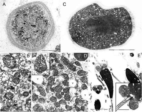
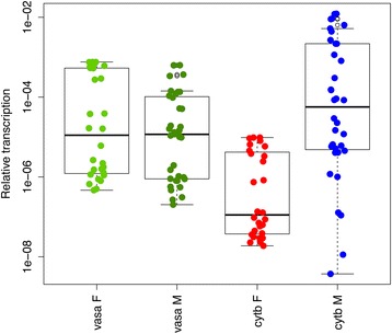
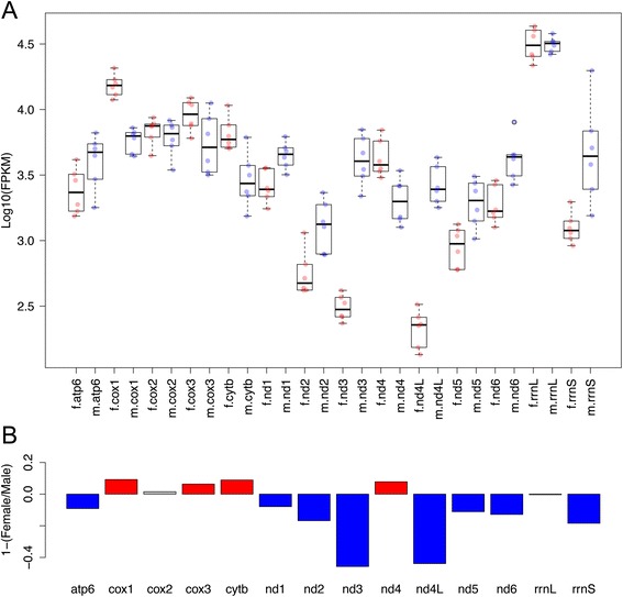
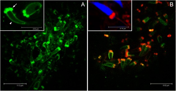
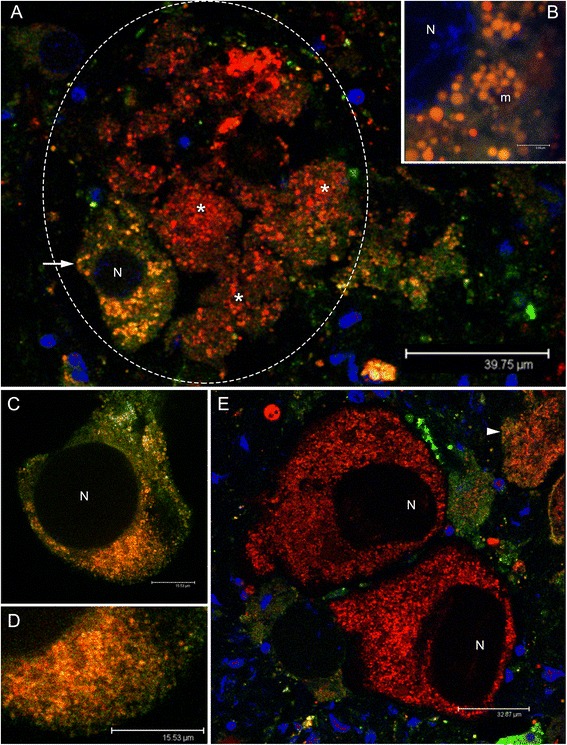
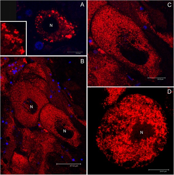
Results: Ultrastructural analysis shows no difference in the conformation of mitochondrial cristae in male and female mature gametes, while mitochondria in immature oocytes exhibit a simpler internal structure. Our data on transcriptional activity in germ line mitochondria show variability between sexes and different developmental stages, but we do not find evidence for transcriptional quiescence of mitochondria. Our observations on mitochondrial membrane potential are consistent with mitochondria being active in both male and female gametes.

Conclusions: Our findings and the literature we discussed may be consistent with the hypothesis that template mitochondria are not functionally silenced, on the contrary their activity might be fundamental for the inheritance mechanism. We think that during gametogenesis, fertilization and embryo development, mitochondria undergo selection for different traits (e.g. replication, membrane potential), increasing the probability of the transmission of functional organelles. In these phases of life cycle, the great reduction in mtDNA copy number per organelle/cell and the stochastic segregation of mtDNA variants would greatly improve the efficiency of selection. When a higher mtDNA copy number per organelle/cell is present, selection on mtDNA deleterious mutants is less effective, due to the buffering effect of wild-type variants. In our opinion, a combination of drift and selection on germ line mtDNA population, might be responsible for the maintenance of viable mitochondrial genetic information through generations, and a mitochondrial activity would be necessary for the selective process.


Figures

Figure 1
Figure 1
Mitochondrial ultrastructure at the transmission electron microscope (TEM). (A) Immature oocyte (scale bar = 10 μm). (B) Immature oocyte mitochondria. Oocytes just starting vitellogenesis show a simple internal structure with few and short mitochondrial cristae (scale bar = 2 μm). Yolk (Y). (C) Mature oocyte (scale bar = 20 μm). (D) Mature oocyte mitochondria. Vitellogenic oocytes show well formed mitochondrial cristae in mitochondria usually measuring less than 600 nm (scale bar = 1 μm). Yolk (Y). (E) Spermatozoa show well formed cristae in mitochondria measuring generally more than 700 nm (scale bar = 2 μm; inset = 0.7 μm). Sperm midpiece containing mitochondria (dashed box); acrosome (a). Inset: spermhead (sh); mitochondria (m); axoneme portion (ax).
Figure 2
Figure 2
Transcript quantification of vasa and cytb genes in developing gonads obtained by RealTime qPCR. The vasa gene shows no differential transcription between males and females, while cytb is significantly more transcribed in males (Wilcoxon Rank-Sum test p-value < 0.001). x-axis: transcription level relative to the reference nuclear gene (18S).
Figure 3
Figure 3
Transcription level of mitochondrial genes in mature gonads obtained by RNA-Seq. (A) Transcription levels expressed in Log10 (FPKM) of mitochondrial genes in females (red dots, “f.” prefix), and males (blue dots, “m.” prefix). (B) Barplot of 1-(Female transcription level/Male transcription level). 0 = no difference; positive values = transcription in females higher than in males; negative values = transcription in males higher than in females; red bars = transcription in females significantly higher than in males (Wilcoxon Rank-Sum test p-value < 0.01); blue bars = transcription in males significantly higher than in females (p-value < 0.01); grey bars = no significant differences. Data from [35].
Figure 4
Figure 4
Mitochondrial inner membrane potential (Δψm) in spermatozoa. (A) The four/five mitochondria of the spermatozoon midpiece (arrow) are visible thanks to Mitotracker Green FM staining. The spermhead (arrowhead) is slightly rimmed by aspecific green staining. (B) The midpiece is strongly red-stained when treated also with Mitotracker Red CMXRos, indicating the presence of a high membrane potential. In the inset the spermhead is stained in blue (TO-PRO 3 nuclear dye).
Figure 5
Figure 5
Mitochondrial inner membrane potential (Δψm) in oocytes (Mitotracker Green FM + Mitotracker Red CMXRos). (A) Female acinus (gonadic unit; dashed oval) containing several oocytes of which one of about 40 μm (arrow), positioned at the periphery of the acinus (acinus wall) and portions of oocytes of higher dimension (asterisks) in the acinus lumen. In the small oocyte the mitochondrial mass appears orange-stained, indicating the presence of a membrane potential (both the color components, Mitotracker Green FM and Mitotracker Red CMXRos, are present). The potential is higher in the biggest oocytes (asterisks), in which the mitochondrial mass is strongly red-stained (Mitotracker Red CMXRos component stronger than the Mitotracker Green FM one). Nuclei of cells around the acinus wall are visible in blue (TO-PRO 3 nuclear dye). (B) Detail of the mitochondrial mass of the small oocyte in (A). Nucleus (N) lightly blue-stained. Mitochondria (m) in orange. (C) Oocyte of about 65 μm in which the mitochondrial mass appears orange-stained, indicating the presence of a membrane potential comparable to that of the 40 μm oocyte in (A, B). The oocyte is starting the pedunculated stage. (D) Detail of the mitochondrial mass of the oocyte in (C). The number of mitochondria appears largely increased in comparison to that of the 40 μm oocyte in (A, B). (E) Two oocytes of about 100 μm at the pedunculated stage shortly before the release into the acinus lumen. The membrane potential is high, as indicated by the strong red staining of the mitochondrial mass (Mitotracker Red CMXRos component stronger than the Mitotracker Green FM one). At the periphery of a close acinus, a small oocyte is visible (arrowhead), showing green-orange staining, indicating the presence of a lower membrane potential in comparison to nearly mature eggs. Nuclei of surrounding cells are blue-stained. Nuclei of oocytes (N) are not always stained since the nuclear labelling is not easily absorbed through the yolk.
Figure 6
Figure 6
Mitochondrial inner membrane potential (Δψm) in oocytes (Mitotracker Red CMXRos). (A) Small oocytes show a less proportion of red-stained mitochondria, than (B) mature eggs (around 100 μm), in which all the mitochondrial mass is red-stained pointing to an overall high membrane potential. (C) Magnification of a mature oocyte inside the acinus (from B). (D) Spawned eggs as well show an overall high mitochondrial membrane potential. Nuclei of cells surrounding the acinus are visible in blue (TO-PRO 3 nuclear dye).

References

    1. Timmis JN, Ayliffe MA, Huang CY, Martin W. Endosymbiotic gene transfer: organelle genomes forge eukaryotic chromosomes. Nat Rev Genet. 2004;5:123–35. - PubMed
    1. Breton S, Milani L, Ghiselli F, Guerra D, Stewart DT, Passamonti M. A resourceful genome: updating the functional repertoire and evolutionary role of animal mitochondrial DNAs. Trends Genet. 2014;30:555-64. - PubMed
    1. Allen JF. Control of gene expression by redox potential and the requirement for chloroplast and mitochondrial genomes. J Theor Biol. 1993;165:609–31. - PubMed
    1. Allen JF. The function of genomes in bioenergetic organelles. Phil Trans R Soc Lond B. 2003;358:19–38. - PMC - PubMed
    1. Lane N, Martin W. The energetics of genome complexity. Nature. 2010;467:929. - PubMed

Publication types

LinkOut - more resources