Case Reports
. 2015 Jul;138(Pt 7):1817-32.
doi: 10.1093/brain/awv117. Epub 2015 May 16.

De novo point mutations in patients diagnosed with ataxic cerebral palsy

Affiliations
Case Reports

De novo point mutations in patients diagnosed with ataxic cerebral palsy

Ricardo Parolin Schnekenberg et al. Brain. 2015 Jul.

Erratum in

  • Corrigendum.
    [No authors listed] [No authors listed] Brain. 2016 Feb;139(Pt 2):e14. doi: 10.1093/brain/awv309. Brain. 2016. PMID: 26912521 Free PMC article. No abstract available.

Abstract

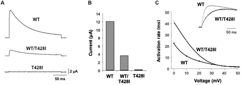
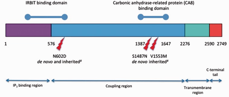
Cerebral palsy is a sporadic disorder with multiple likely aetiologies, but frequently considered to be caused by birth asphyxia. Genetic investigations are rarely performed in patients with cerebral palsy and there is little proven evidence of genetic causes. As part of a large project investigating children with ataxia, we identified four patients in our cohort with a diagnosis of ataxic cerebral palsy. They were investigated using either targeted next generation sequencing or trio-based exome sequencing and were found to have mutations in three different genes, KCNC3, ITPR1 and SPTBN2. All the mutations were de novo and associated with increased paternal age. The mutations were shown to be pathogenic using a combination of bioinformatics analysis and in vitro model systems. This work is the first to report that the ataxic subtype of cerebral palsy can be caused by de novo dominant point mutations, which explains the sporadic nature of these cases. We conclude that at least some subtypes of cerebral palsy may be caused by de novo genetic mutations and patients with a clinical diagnosis of cerebral palsy should be genetically investigated before causation is ascribed to perinatal asphyxia or other aetiologies.

Keywords: ataxia; cerebral palsy; de novo; intellectual disability.


Figures

None
Cerebral palsy is commonly attributed to perinatal asphyxia. However, Schnekenberg et al. describe here four individuals with ataxic cerebral palsy likely due to de novo dominant mutations associated with increased paternal age. Therefore, patients with cerebral palsy should be investigated for genetic causes before the disorder is ascribed to asphyxia.
Figure 1
Figure 1
MRI of the brains of Cases 2, 3 and 4. (A and B) Cases 2 and 3 showing a normal brain MRI. (C) Case 4 brain MRI shows a small cerebellum, with increased spacing of the cerebellar folia and an enlarged fourth ventricle. This was reported to be cerebellar atrophy, rather than hypoplasia.
Figure 2
Figure 2
Confirmation of parentage in Cases 1–4. (A) Sequences of rare SNPs in parents and affected of Case 1 showing consistency with parentage: genes, variant and genomic location (hg19) are shown. (B) The non-reference discordance rate (NDR) over 86 000 exonic Hapmap SNPs for Cases 2, 3 and 4. Related individuals show lower discordance (yellow) than unrelated individuals (blue/purple). This analysis confirms that the probands in the study are genetically related to both parents and that parents are not genetically related to each other.
Figure 3
Figure 3
A novel de novo mutation predicted to affect the S4 voltage-sensor of Kv3.3. (A) The high degree of amino acid conservation (asterisk) in the voltage-sensor S4 helix and S4-S4 linker region of human Kv3.3 and related species. This region is also highly conserved in the paralogous channels KCNC1 (Kv3.1) and KCNC2 (Kv3.2). Threonine 428 in KCNC3 (Kv3.3) is highlighted in grey and is absolutely conserved between species. (B) Sanger sequencing of the patient and parents to show that the heterozygous mutation is de novo. (C) A structural model of this region in Kv3.3 with the predicted location of the T428I mutation. The conserved voltage-sensing arginine and lysine residues are also shown.
Figure 4
Figure 4
The T428I mutation affects the functional properties of Kv3.3. (A) Representative current traces recorded from homomeric wild-type (WT), homomeric mutant (T428I) and heteromeric (WT/T428I) Kv3.3 channels. The mutant channel exhibits a severe dominant-negative, loss-of-function phenotype. (B) Quantifies this dominant negative effect in heteromeric (WT/T428I) channels that mimic the heterozygous state. (C) Demonstrates that the residual current in the heterozygous state (WT/T428I) has markedly altered gating properties with much slower rates of activation across a wide range of voltages. The inset panel compares representative traces for wild-type and heteromeric (WT/T428I) channels recorded at +60 mV.
Figure 5
Figure 5
Location of ITPR1 missense mutations in functional domains of the IP3R protein. De novo N587D and S1487N are described in this paper. Inherited mutations (hash) are reported previously (Huang et al., 2012).
Figure 6
Figure 6
Peak sodium current enhanced less by R480W than wild-type β-III spectrin. (A) Sodium current traces from representative cells evoked with a series of 50 ms depolorizations from a holding potential of −90 mV to potentials ranging from −80 to +20 mV in 10 mV increments (stimulus protocol shown at bottom). (B) Sodium current peak at −10 mV normalized to control cells cultured at same time. (C) Current-voltage relationships for control, wild-type (WT) and R480W with current amplitude normalized to peak value. All data are presented as the mean ± SEM (n = 5–9 cells from each of three independent cultures; P < 0.05).

References

    1. Adzhubei IA, Schmidt S, Peshkin L, Ramensky VE, Gerasimova A, Bork P, et al. A method and server for predicting damaging missense mutations. Nat Methods 2010; 7: 248–9. - PMC - PubMed
    1. Allen AS, Berkovic SF, Cossette P, Delanty N, Dlugos D, Eichler EE, et al. De novo mutations in epileptic encephalopathies. Nature 2013; 501: 217–21. - PMC - PubMed
    1. Ando H, Mizutani A, Kiefer H, Tsuzurugi D, Michikawa T, Mikoshiba K. IRBIT suppresses IP3 receptor activity by competing with IP3 for the common binding site on the IP3 receptor. Mol Cell 2006; 22: 795–806. - PubMed
    1. Bauer P, Schols L, Riess O. Spectrin mutations in spinocerebellar ataxia (SCA). Bioessays 2006; 28: 785–7. - PubMed
    1. Bourassa CV, Meijer IA, Merner ND, Grewal KK, Stefanelli MG, Hodgkinson K, et al. VAMP1 mutation causes dominant hereditary spastic ataxia in Newfoundland families. Am J Hum Genet 2012; 91: 548–52. - PMC - PubMed

Publication types

Supplementary concepts