. 2015 Sep;21(17-18):2315-22.
doi: 10.1089/ten.TEA.2014.0622. Epub 2015 Jul 13.

Intramyocardial Delivery of Notch Ligand-Containing Hydrogels Improves Cardiac Function and Angiogenesis Following Infarction

Affiliations

Intramyocardial Delivery of Notch Ligand-Containing Hydrogels Improves Cardiac Function and Angiogenesis Following Infarction

Archana V Boopathy et al. Tissue Eng Part A. 2015 Sep.

Abstract

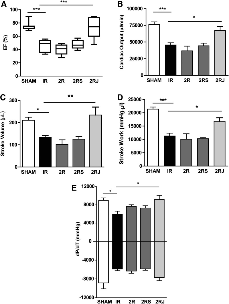
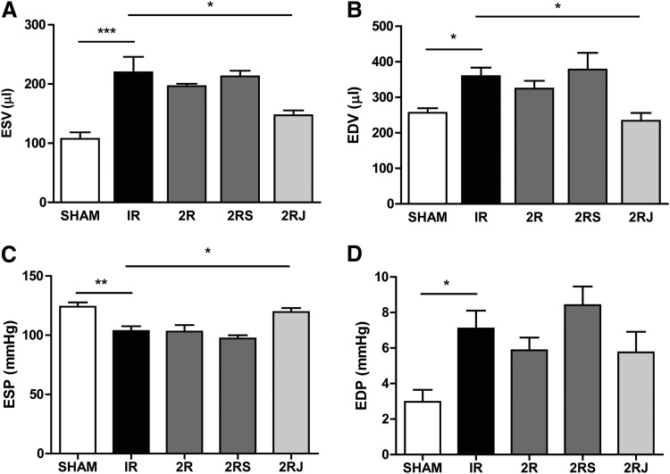
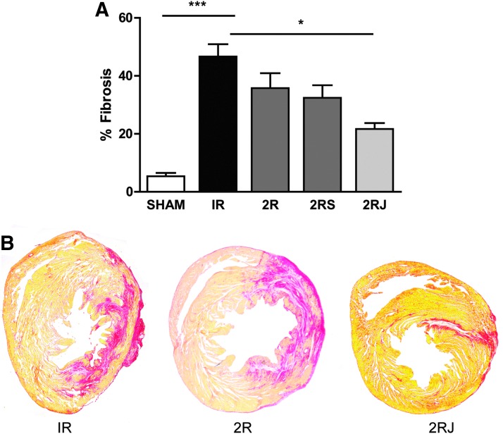
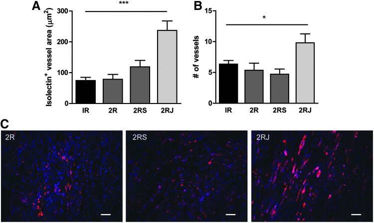
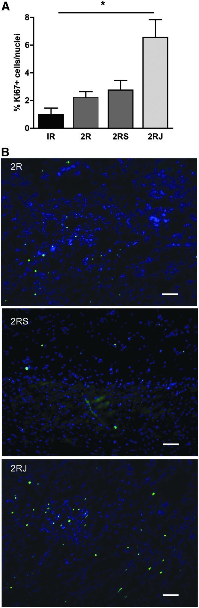
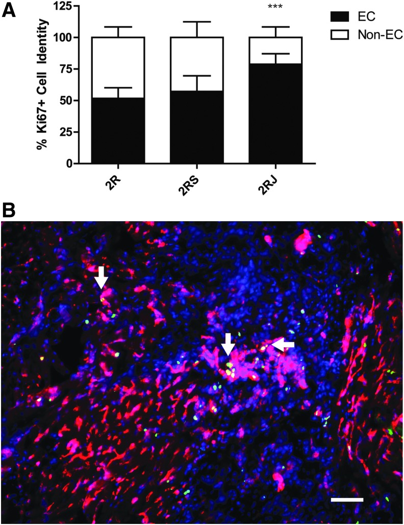
Myocardial infarction (MI) is the leading cause of death worldwide. Notch1 signaling plays a critical role in cardiac development, in survival, cardiogenic lineage commitment, differentiation of cardiac stem/progenitor cells, and in regenerative responses following myocardial injury. The objective of this study was the evaluation of the therapeutic effect of delivering the Notch ligand-containing hydrogels in a rat model of MI. Self-assembling peptide (SAP) hydrogels were functionalized with a peptide mimic of the Notch1 ligand Jagged1 (RJ). In rats subjected to experimental MI, delivery of RJ-containing hydrogel to the infarcted heart resulted in improvement in cardiac function back to sham-operated levels. A significant decrease in fibrosis and an increase in the endothelial vessel area and Ki67 expression were also observed in rats treated with the RJ hydrogels compared to untreated rats or rats treated with unmodified or scrambled peptide hydrogels. This study demonstrates the functional benefit of Notch1-activating peptide delivered in SAP hydrogels for cardiac repair.


Figures

<b>FIG. 1.</b>
FIG. 1.
Delivery of 2RJ hydrogel improves cardiac function following MI. Pressure–volume hemodynamic measurements of (A) ejection fraction (EF%), (B) cardiac output, (C) stroke volume, (D) stroke work, (E)±dP/dT; n≥6/group, *p<0.05, **p<0.01, ***p<0.001, one-way ANOVA followed by Tukey's post-test. MI, myocardial infarction; 2R, 2% RAD hydrogel; 2RS, 2% RAD hydrogel with scrambled peptide; 2RJ, 2% RAD hydrogel with Jagged peptide.
<b>FIG. 2.</b>
FIG. 2.
Delivery of 2RJ hydrogel improves left ventricular morphology following MI. Pressure–volume hemodynamic measurements of (A) ESV, (B) EDV, (C) ESP, and (D) EDP. *p<0.05, **p<0.01, n≥5/group. One-way ANOVA followed by Tukey's post-test. ESV, end-systolic volume; EDV, end-diastolic volume; ESP, end-systolic pressure; EDP, end-diastolic pressure.
<b>FIG. 3.</b>
FIG. 3.
Delivery of 2RJ hydrogel decreases fibrosis. (A) Quantification of % fibrosis in rat hearts on day 21 shows a significant increase in fibrosis following 30 min of IR (black bar). Administration of 2RJ hydrogel attenuated this increase; ***p<0.001 versus sham, *p<0.05 versus IR, n≥5 per group. One-way ANOVA followed by Tukey's post-test. (B) Representative images of heart sections stained with picrosirius red and stitched together. IR, ischemia–reperfusion.
<b>FIG. 4.</b>
FIG. 4.
Delivery of 2RJ hydrogel increases endothelial vessel area. Quantification of endothelial vessel area (A) and number of vessels (B) in rat hearts on day 21 by isolectin staining shows a significant increase in both measurements in infarcted rats treated with 2RJ hydrogel. ***p<0.001 and *p<0.05 versus IR, n=4−7 per group. (C) Representative images of heart sections stained with isolectin (red) and DAPI (blue). Scale bar=50 μm.
<b>FIG. 5.</b>
FIG. 5.
Delivery of 2RJ hydrogel increases Ki67 expression. (A) Quantification of Ki67+ cells in rat hearts on day 21 shows an increase in Ki67+ cell number in infarcted rats treated with 2RJ hydrogel. *p<0.05, n=4 per group. (B) Representative images of Ki67 staining in the myocardium. Blue, DAPI; green, Ki67 nuclei. Scale bar=50 μm.
<b>FIG. 6.</b>
FIG. 6.
Ki67+ cells in RJ are mostly endothelial cells. (A) Quantification of double-positive Ki67 and isolectin cells in each group shows a significant change in cell lineage in 2RJ-treated rats. ***p<0.001 versus IR, n=4 per group. One-way ANOVA followed by Tukey's post-test. (B) Representative image from a 2RJ-treated rat heart showing isolectin (red), Ki67 (green), and DAPI (blue). Arrows denote areas of double staining. Scale bar=50 μm.

References

    1. Go A.S., Mozaffarian D., Roger V.L., Benjamin E.J., Berry J.D., Blaha M.J., et al. . Heart disease and stroke statistics—2014 update: a report from the American Heart Association. Circulation 129, e28, 2014 - PMC - PubMed
    1. Anversa P. Myocyte apoptosis and heart failure. Eur Heart J 19, 359, 1998 - PubMed
    1. Weisman H.F., and Healy B. Myocardial infarct expansion, infarct extension, and reinfarction: pathophysiologic concepts. Prog Cardiovasc Dis 30, 73, 1987 - PubMed
    1. Enomoto Y., Gorman J.H., 3rd, Moainie S.L., Jackson B.M., Parish L.M., Plappert T., et al. . Early ventricular restraint after myocardial infarction: extent of the wrap determines the outcome of remodeling. Ann Thorac Surg 79, 881, 2005 - PubMed
    1. Christman K.L., Fok H.H., Sievers R.E., Fang Q., and Lee R.J. Fibrin glue alone and skeletal myoblasts in a fibrin scaffold preserve cardiac function after myocardial infarction. Tissue Eng 10, 403, 2004 - PubMed

Publication types

MeSH terms