. 2015 Jun 4;96(6):894-912.
doi: 10.1016/j.ajhg.2015.04.011. Epub 2015 May 14.

Concurrent whole-genome haplotyping and copy-number profiling of single cells

Affiliations

Concurrent whole-genome haplotyping and copy-number profiling of single cells

Masoud Zamani Esteki et al. Am J Hum Genet. .

Abstract

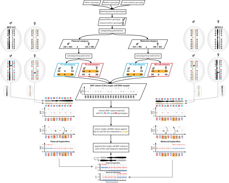
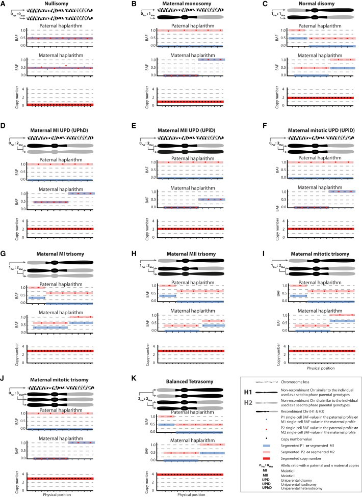
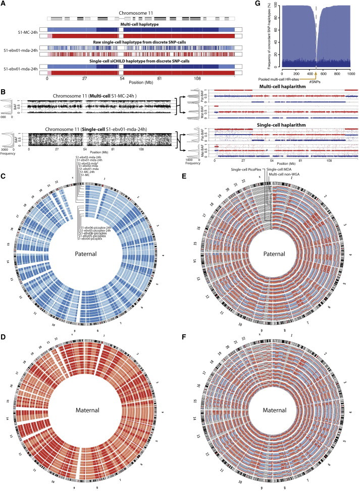
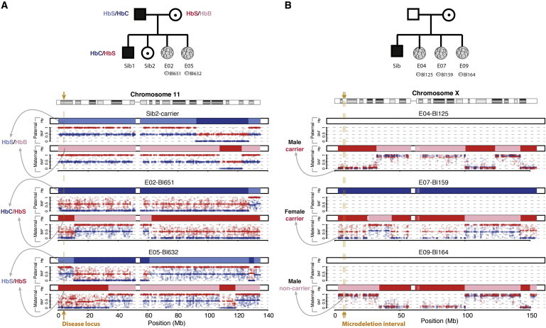
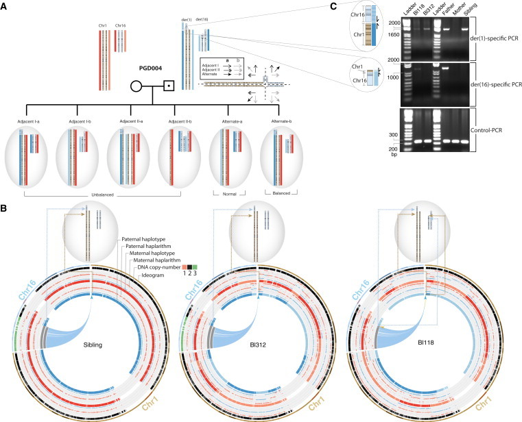
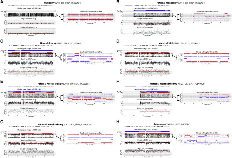
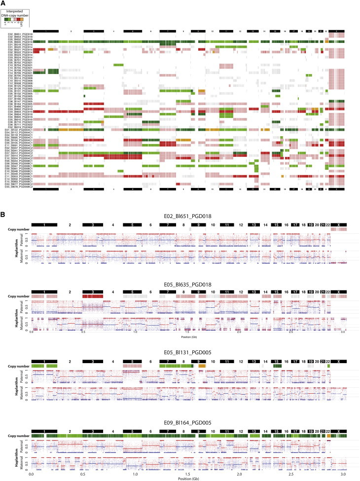
Methods for haplotyping and DNA copy-number typing of single cells are paramount for studying genomic heterogeneity and enabling genetic diagnosis. Before analyzing the DNA of a single cell by microarray or next-generation sequencing, a whole-genome amplification (WGA) process is required, but it substantially distorts the frequency and composition of the cell's alleles. As a consequence, haplotyping methods suffer from error-prone discrete SNP genotypes (AA, AB, BB) and DNA copy-number profiling remains difficult because true DNA copy-number aberrations have to be discriminated from WGA artifacts. Here, we developed a single-cell genome analysis method that reconstructs genome-wide haplotype architectures as well as the copy-number and segregational origin of those haplotypes by employing phased parental genotypes and deciphering WGA-distorted SNP B-allele fractions via a process we coin haplarithmisis. We demonstrate that the method can be applied as a generic method for preimplantation genetic diagnosis on single cells biopsied from human embryos, enabling diagnosis of disease alleles genome wide as well as numerical and structural chromosomal anomalies. Moreover, meiotic segregation errors can be distinguished from mitotic ones.


Figures

Figure 1
Figure 1
The Principles of Haplarithmisis The sequence of actions applied for deciphering haplotypes and concomitantly copy number, parent-of-origin, and segregational origin information from single-cell SNP BAF values. The figure illustrates how maternal and paternal haplarithm plots arise for a cell that contains a normal disomy with one homologous recombination on each inherited chromosome. Parental homologs 1 and 2 (H1 and H2, respectively) are defined on the basis of their phased genotype. Pairwise breakpoints in the segmented M1 and M2 single-cell SNP BAF values pinpoint maternal homologous recombination sites, likewise for P1 and P2 in the paternal haplarithm plot. Additionally, the positioning of M1-M2 and P1-P2 segments is expected to be at 0, 0.5, or 1 on the y axis for a disomic copy number.
Figure 2
Figure 2
Unique Haplarithm Patterns for Different Chromosomal Anomalies The segmented M1 and M2 single-cell BAF values, as well as the distance between M1 and M2 values, changes according to the copy-number anomaly, the affected parental allele, and the meiotic I (MI), meiotic II (MII), or mitotic origin of the anomaly. Pairwise P1-P2 and M1-M2 breakpoints in the haplarithm profiles delineate sites of homologous recombination (i.e., the parity feature of haplarithmisis). Separately, the distance between P1 and P2 values (dPat) changes in a reciprocal manner with the M1-M2 distance (dMat) denoting copy number, parent-of-origin, and segregational origin of haplotypes (i.e., the reciprocity feature of haplarithmisis). Shown are expected haplarithm patterns for a nullisomy (A), a maternal monosomy (B), a normal disomy (C), a maternal MI UPD (D), a maternal MII UPD (E), a maternal mitotic UPD (F), a maternal MI trisomy (G), a maternal MII trisomy (H), a maternal mitotic trisomy (I), a maternal mitotic trisomy with three identical chromosomes (J), and a balanced tetrasomy (K).
Figure 3
Figure 3
Whole-Genome Single-Cell Haplotyping (A) Multi-cell and single-cell haplotypes of a disomic chromosome using discrete SNP calls before and after siCHILD analysis. (B) Multi-cell and single-cell haplotypes of the same chromosome using continuous SNP BAF values before and after siCHILD’s haplarithmisis of the same samples. Histograms and density plots of the SNP BAF profiles before and after haplarithmisis are juxtaposed. (C and D) Genome-wide haplotypes obtained from the discrete SNP calls via siCHILD. (E and F) The genome-wide haplarithm profiles of the same samples derived from continuous SNP BAF values. (G) Concordance of single-cell SNP-haplotype calls (via discrete genotypes) with the reference haplotype of the matching multi-cell DNA samples.
Figure 4
Figure 4
siCHILD-Based PGD for Mendelian Disorders Applying siCHILD on single blastomeres, we traced the inheritance of parental disease variants in human IVF embryos. (A) In a PGD case subject segregating mutant HbS and HbC alleles underlying the autosomal-recessive sickle-cell anemia. (B) In a PGD case subject with an X-linked Xp22.31 microdeletion recessive disorder.
Figure 5
Figure 5
siCHILD-Based PGD for Translocation Carriers (A) The main possible modes of inheritance of the derivative chromosomes of a reciprocal translocation present in a carrier father to his IVF embryos are depicted. Importantly, by determining the haplotypes flanking the translocation breakpoints, the inheritance of the normal and derivative chromosomes involved in a parental reciprocal translocation can be traced to an embryo. (B) Applying siCHILD on single blastomeres, we traced the inheritance of the normal and derivative chromosomes of a paternal balanced reciprocal translocation t(1;16)(p36;p12) to his embryos after IVF. Breakpoint flanking haplotypes indicated the inheritance of der(1) and a normal chromosome 16 in cell Bl312, as well as of der(1) and der(16) chromosomes in cell Bl118. (C) PCR-based validation of the inherited (derivative) chromosomes via primers designed after single-cell paired-end sequencing.
Figure 6
Figure 6
Single-Cell Copy-Number Analysis Supervised by Haplarithmisis: Full Chromosome Anomalies Different aneuploidies, detected by SNP-array and single-cell sequencing, are authenticated by different characteristic haplarithm patterns. In addition, the parental haplarithm profiles disclose the haplotype-specific copy-number states of the chromosomes in the cells and reveal the parental and meiotic/mitotic origin of the chromosomal anomaly. We show (A) a nullisomy (i.e., 0Pat:0Mat allelic ratio), (B) a paternal monosomy (1Pat:0Mat), (C) a normal disomy (1Pat:1Mat), (D) a mitotic maternal UPD (0Pat:2Mat), (E) a paternal trisomy (2Pat:1Mat), (F) a meiotic maternal trisomy (1Pat:2Mat), (G) a maternal trisomy (0Pat:3Mat), and (H) a tetrasomy (2Pat:2Mat).
Figure 7
Figure 7
Aneuploidy Screening of All 60 Single Blastomeres (A) Genome-wide copy-number maps of the single blastomeres. Aberrant logR segments (>0.15 or <−0.3) corroborated by a distinctive haplarithm pattern are depicted. Aberrant logR segments not corroborated by a typical haplarithm pattern are depicted in gray. For cells analyzed in the framework of translocation PGD case subjects, DNA imbalances smaller than 3 Mb corroborated by the haplotype of the derivative chromosome of the parental reciprocal translocation are depicted as well. (B) Genome-wide copy-number, paternal, and maternal haplarithm profiles of four single blastomeres (for detailed parental haplarithm patterns for each cell, see Figure S14).

References

    1. Baudat F., Imai Y., de Massy B. Meiotic recombination in mammals: localization and regulation. Nat. Rev. Genet. 2013;14:794–806. - PubMed
    1. Tewhey R., Bansal V., Torkamani A., Topol E.J., Schork N.J. The importance of phase information for human genomics. Nat. Rev. Genet. 2011;12:215–223. - PMC - PubMed
    1. Vanneste E., Voet T., Le Caignec C., Ampe M., Konings P., Melotte C., Debrock S., Amyere M., Vikkula M., Schuit F. Chromosome instability is common in human cleavage-stage embryos. Nat. Med. 2009;15:577–583. - PubMed
    1. van Echten-Arends J., Mastenbroek S., Sikkema-Raddatz B., Korevaar J.C., Heineman M.J., van der Veen F., Repping S. Chromosomal mosaicism in human preimplantation embryos: a systematic review. Hum. Reprod. Update. 2011;17:620–627. - PubMed
    1. Voet T., Vanneste E., Vermeesch J.R. The human cleavage stage embryo is a cradle of chromosomal rearrangements. Cytogenet. Genome Res. 2011;133:160–168. - PubMed

Publication types

Associated data